Article Text

Abstract
Leptospirosisis a zoonosis caused by spirochaetes from the species Leptospira. The more severe form of leptospirosis, known as Weil’s disease, is characterised by the triad of jaundice, renal impairment and haemorrhages. Pulmonary involvement occurs in 20%–70% of the patients, with severity ranging from non-productive cough to respiratory failure mainly due to pulmonary haemorrhage. Recognition of Weil’s disease in patients presenting with pulmonary symptoms can be difficult. This case illustrates a classic case of pulmonary haemorrhagic involvement in Weil’s disease.
- respiratory medicine
- infectious diseases
This is an open access article distributed in accordance with the Creative Commons Attribution Non Commercial (CC BY-NC 4.0) license, which permits others to distribute, remix, adapt, build upon this work non-commercially, and license their derivative works on different terms, provided the original work is properly cited and the use is non-commercial. See: http://creativecommons.org/licenses/by-nc/4.0/.
Statistics from Altmetric.com
Background
Leptospirosis is a zoonosis caused by spirochaetes from the species Leptospira. Host animals carry the leptospires in their kidneys and shed the pathogen in their urine.1 Infections result from direct or indirect contact with infected animals, from which the brown rat serves as the most important reservoir for human infection.2 Costa et al estimated in a recent review that the worldwide incidence of leptospirosis is approximately 1.03 million cases and that almost 60 000 deaths annually occur due to leptospirosis.3 Relatively unknown are the high rates of pulmonary involvement in patients with leptospirosis. In the literature, pulmonary leptospirosis is mostly described as a separate disease entity, or as a symptom associated with worse outcome. Early recognition of all symptoms of the disease and initiation of treatment is crucial in order to decrease the risk of severity and/or fatality. This patient presents with the characteristic triad of symptoms and pulmonary haemorrhage.
Case presentation
A 26-year-old previously healthy man presented to the emergency department. He returned 1 week earlier from a holiday in Jamaica. He had swam in fresh water, while dead rats were floating next to him. Since 5 days he was experiencing fever, nausea, muscle pain in the legs, jaundice, oliguria, headaches and haemoptysis. Physical examination revealed a sick young man with jaundice, conjunctival suffusion and tenderness of the epigastric region. No other abnormalities were observed.
Investigations
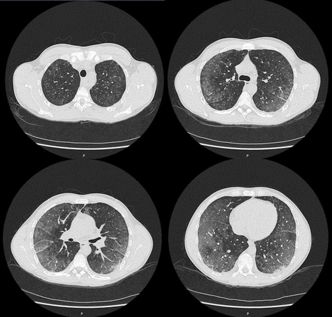
Blood tests showed a serum creatinine level of 631 µmol/L (reference 59–104), urea of 17.6 mmol/L (reference 2.5–7.5), sodium of 136 mmol/L (reference 136–144), potassium of 3.3 mmol/L (reference 3.5–5), haemoglobin of 7.6 mmol/L (reference 5.8–11), thrombocytopenia of 21×109/L (reference 150–400), leukocytosis of 13.6×10⁹/L (reference 4.3–10.0), haptoglobin of 3.9 g/L (reference 0.3–2.0 g/L), lactate dehydrogenase of 244 U/L (reference <248), creatine kinase of 620 U/L (reference <200) and mildly elevated liver enzymes as alanine aminotransferase was 44 U/L (reference <50), aspartate aminotransferase 67 U/L (reference <45), alkaline phosphatase 152 U/L (reference <125). Total bilirubin was 408 µmol/L (reference 3.4–20.8) with a conjugated bilirubin of 332 µmol/L. Gamma-glutamyltransferase could not be measured due to the high level of bilirubin. Anti-neutrophil cytoplasmic antibodies were negative. A peripheral smear showed 80% neutrophils, 5% lymphocytes and 14% monocytes, target cells, lymphocytes bigger than normal and lymphocytes pressed between erythrocytes were also seen. Urine examination showed a pH of 6.0 (reference 4.5–8.0), erythrocytes positive (1+), leucocytes negative, nitrite negative, albumin/creatinine ratio 782.0 mg/mmol (reference <2.5) and microalbumin 3003.0 mg/L (reference <20 mg/L). Investigations for hantavirus, hepatitis A, B and C were performed and turned out negative. An ultrasound of the abdomen revealed no abnormalities of liver, gall bladder and kidneys. A chest X-ray at presentation showed an interstitial lung pattern, consisting of nodular and reticular opacifications (figure 1). In addition, high-resolution CT (HRCT) of the thorax was performed (figure 2). CT showed extensive ground-glass opacities with more centrilobular located aberrations in the upper lung fields, and predominantly peripheral abnormalities in the lower lung fields. These radiological findings combined with haemoptysis were highly suggestive for alveolar haemorrhage.

A chest X-ray showed an interstitial lung pattern consisting of nodular and reticular opacifications.
CT showed extensive ground-glass opacities with more centrilobular located aberrations in the upper lung fields, and predominantly peripheral abnormalities in the lower lung fields.
Differential diagnosis
The differential diagnostic included leptospirosis, malaria, hantavirus, dengue and chikungunya. In the absence of haemolysis (normal haemoglobin and haptoglobin), malaria was less likely.4 Hantavirus is a rodent-borne viral disease. It usually presents itself with haemorrhagic fever, renal insufficiency and/or pulmonary symptoms. Jaundice due to a high level of bilirubin is not a common feature.5 Kidney and liver failure are seen in dengue shock syndrome as a result of intravascular volume depletion due to plasma leakage.6 Our patient was haemodynamically stable, not compatible with dengue. One of the characteristic features of chikungunya is polyarthralgia, which was not present in our patient.7 The modified Faine’s criteria were used for estimating the change of leptospirosis based on clinical history (part A) and epidemiological history (part B) supported by laboratory findings (part C). A score of 19 on part A and 6 on part B (points for part C could not be given because laboratory results were not available yet) made a total score of 25 on the modified Faine’s criteria. A score of 26 or more on part A or part A+B makes leptospirosis a presumptive diagnosis; a score between 20 and 25 is suggestive for leptospirosis.8
Treatment
As leptospirosis was suspected, the patient was directly started on intravenous penicillin (12 million units daily). Intravenous fluid suppletion was started with 3 litres of 0.9% sodium chloride. Dialysis was considered; however, in the absence of uraemic symptoms (eg, encephalopathy), signs of volume overload or electrolyte abnormalities, renal replacement therapy was not initiated.
Outcome and follow-up
The diagnosis of leptospirosis was confirmed after 5 days by PCR of the serum. The patient responded well to the antibiotic treatment and quickly recovered. Antibiotic treatment was continued for 7 days. The intravenous fluid suppletion was reduced and stopped the day before discharge. Platelet count and renal function improved after initiation of therapy and were normalised at the time of discharge. Bilirubin and liver enzyme levels showed a declining trend. Haemoptysis did not increase and was no longer reported by the patient 2 weeks after discharge.
Discussion
Most leptospirosis infections are mild and cause influenza-like illness.2 The more severe form of leptospirosis, known as Weil’s disease, is characterised by the triad of jaundice, renal impairment and haemorrhages, and has a high mortality rate of 5%–15%.9 10 The classic course of leptospirosis is characterised by a biphasic pattern, starting with a flu-like bacteraemic phase in which the leptospires circulate in the bloodstream. This acute phase is followed by an immune phase, in which the leptospiral toxins and immune-mediated responds can cause a broad spectrum of complications as shock, renal failure and massive pulmonary haemorrhage.11 Pulmonary involvement occurs in 20%–70% of patients with leptospirosis and sometimes is the presenting symptom.12 Its severity ranges from a non-productive cough to respiratory failure due to extensive pulmonary haemorrhage and the formation of hyaline membranes.13 After onset, pulmonary symptoms can be rapidly progressive and patients can die in less than 48 hours as a result of respiratory failure.14 The haemorrhagic form has a mortality rate of up to 60%.15–18 Chest radiography can already show abnormalities as early as 24 hours after the start of pulmonary symptoms. Bilateral diffuse air space disease is the most common finding on abnormal chest X-ray.19 Characteristic radiological findings of pulmonary involvement in leptospirosis on CT are bilateral ground-glass opacities mainly in the peripheral and lower long zones. Areas of consolidation, air-space nodules and small pleural effusions are other characteristics that can be seen.20 Pulmonary involvement is one of the predictors associated with worse outcome. Therefore, it is very important to recognise this involvement as potentially presenting symptom of Weil’s disease.9 This case report illustrates characteristic findings of pulmonary haemorrhage on HRCT of the lungs, which could be helpful in future diagnosis. In a retrospective case–control study, early initiation of antibiotics decreased the risk of developing severe disease.21 Our patient demonstrates that early initiation of antibiotics in patients already presenting with a severe form of leptospirosis can be effective in preventing progression of the disease. In summary, this case illustrates a severe case of Weil’s disease including pulmonary haemorrhage with full recovery after initiation of treatment in early state of disease.
Learning points
Pulmonary involvement is a frequently seen symptom in leptospirosis, with severity ranging from non-productive cough to pulmonary haemorrhage and respiratory failure.
Radiological findings of pulmonary involvement in leptospirosis on CT are bilateral ground-glass opacities mainly in the peripheral and lower long zones.
Full recovery is seen when treated appropriately with penicillin.
References
Footnotes
Contributors EB and SH wrote the manuscript with support from JEB. LO helped supervise the project. All authors took care of the patient during his hospital admission and contributed to the final manuscript.
Funding The authors have not declared a specific grant for this research from any funding agency in the public, commercial or not-for-profit sectors.
Competing interests None declared.
Provenance and peer review Not commissioned; externally peer reviewed.
Patient consent for publication Obtained.