Article Text

Abstract
Renal transplant recipients are prone to opportunistic infections due to iatrogenic immunosuppression. Infectious esophagitis can present as an opportunistic infection in the post-transplant period. Common pathogens are candida, herpes simplex virus (HSV) and cytomegalovirus (CMV). Having a dual infection is uncommon and the diagnoses can be missed at initial presentation. Our patient, a 29-year-old African-American woman, status post deceased-donor-kidney transplant presented with difficulty and pain in swallowing with clinical features suggestive of candida esophagitis, confirmed by fungal culture. She did not get better with antifungal treatment. On further testing, the patient was found to have HSV-2 infection of the oesophagus as well. She received both fluconazole as well as acyclovir that lead to complete resolution of her symptoms. In the right clinical setting, esophagitis can be caused by more than one organism present at the same time and a high level of suspicion is warranted.
- renal transplantation
- hepatitis and other gi infections
- infections
- endoscopy
- infection (gastroenterology)
This is an open access article distributed in accordance with the Creative Commons Attribution Non Commercial (CC BY-NC 4.0) license, which permits others to distribute, remix, adapt, build upon this work non-commercially, and license their derivative works on different terms, provided the original work is properly cited and the use is non-commercial. See: http://creativecommons.org/licenses/by-nc/4.0/
Statistics from Altmetric.com
- renal transplantation
- hepatitis and other gi infections
- infections
- endoscopy
- infection (gastroenterology)
Background
Renal transplant patients are prone to infectious esophagitis primarily because of their immunosuppressed status. Candida, herpes simplex virus (HSV) and cytomegalovirus (CMV) are the usual causative agents in such patients, but having esophagitis due to two organisms simultaneously is uncommon and can be missed at the time of initial presentation, leading to clinical complications. A dual pathogen esophagitis is usually not suspected. Also, clinical features of one causative agent may dominate over the other. Missing such diagnoses in immunosuppressed renal transplant patients can lead to worse clinical outcomes.
Case presentation
A 29-year-old African-American woman received a deceased-donor -kidney transplant in September 2017. She was induced with pulse steroids and thymoglobulin. Her renal graft function was stable on maintenance immunosuppression with mycophenolic acid 1000 mg two times per day, prednisone 5 mg daily and tacrolimus with a goal level of 6–8 ng/mL. Tacrolimus level remained within the therapeutic range. She also received valgancyclovir, nystatin and Bactrim for 3 months post-transplantation as prophylaxis for opportunistic infections. The patient had a negative HSV-1 and HSV-2 serology prior to the transplant. Donor HSV-1 and HSV-2 status was not known. The patient had an episode of biopsy-proven, borderline, acute cellular rejection in the second month after transplant that responded well to pulse steroids with normalisation of renal function.
Nine months after her kidney transplantation she presented with sore throat, dysphagia, odynophagia and fever. Her physical examination was significant for oropharyngeal thrush, white tonsillar exudates, pharyngeal erythema and enlarged palatine tonsils. Blood work showed leukocytosis and acute kidney injury (AKI). She was admitted to the hospital for intravenous fluids and intravenous fluconazole therapy for severe oropharyngeal candidiasis and high suspicion of esophageal candidiasis. After 48 hours of intravenous antifungal treatment, the patient had improvement of symptoms and was discharged home on oral fluconazole and clindamycin. Clindamycin was empirically prescribed for the treatment of any underlying tonsillitis.
Three days post discharge, the patient was readmitted in a septic state, with worse odynophagia, tachycardia, high-grade fever and worsening leukocytosis. The patient appeared ill. Physical examination again demonstrated white plaques on the tongue, palate and tonsils with bilateral lingual and palatine tonsillar swelling (figure 1). The white plaques on the tongue were amenable to scraping. A CT scan of the neck was obtained showing swollen lingual and palatine tonsils with narrowing of the pharynx and bilateral level 2A and 2B cervical lymphadenopathy. It did not show any drainable abscess. The patient was started on empiric intravenous antibiotics and intravenous fluconazole was restarted. The patient’s fever and tachycardia improved but she continued to have discomfort and pain in her throat while swallowing as well as in her chest. Throat culture was positive for candida albicans and negative for any bacterial growth. Blood cultures were negative. Rapid streptococcal antigen test, Monospot test, Epstein bar virus PCR and CMV PCR were also negative.
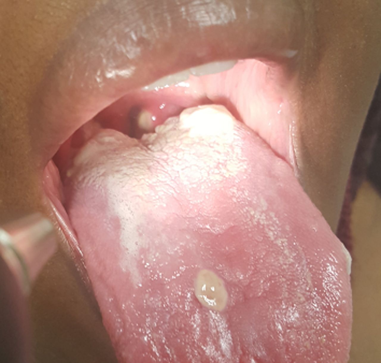
White plaques on tongue and tonsils with bilateral lingual and palatine tonsillar swelling.
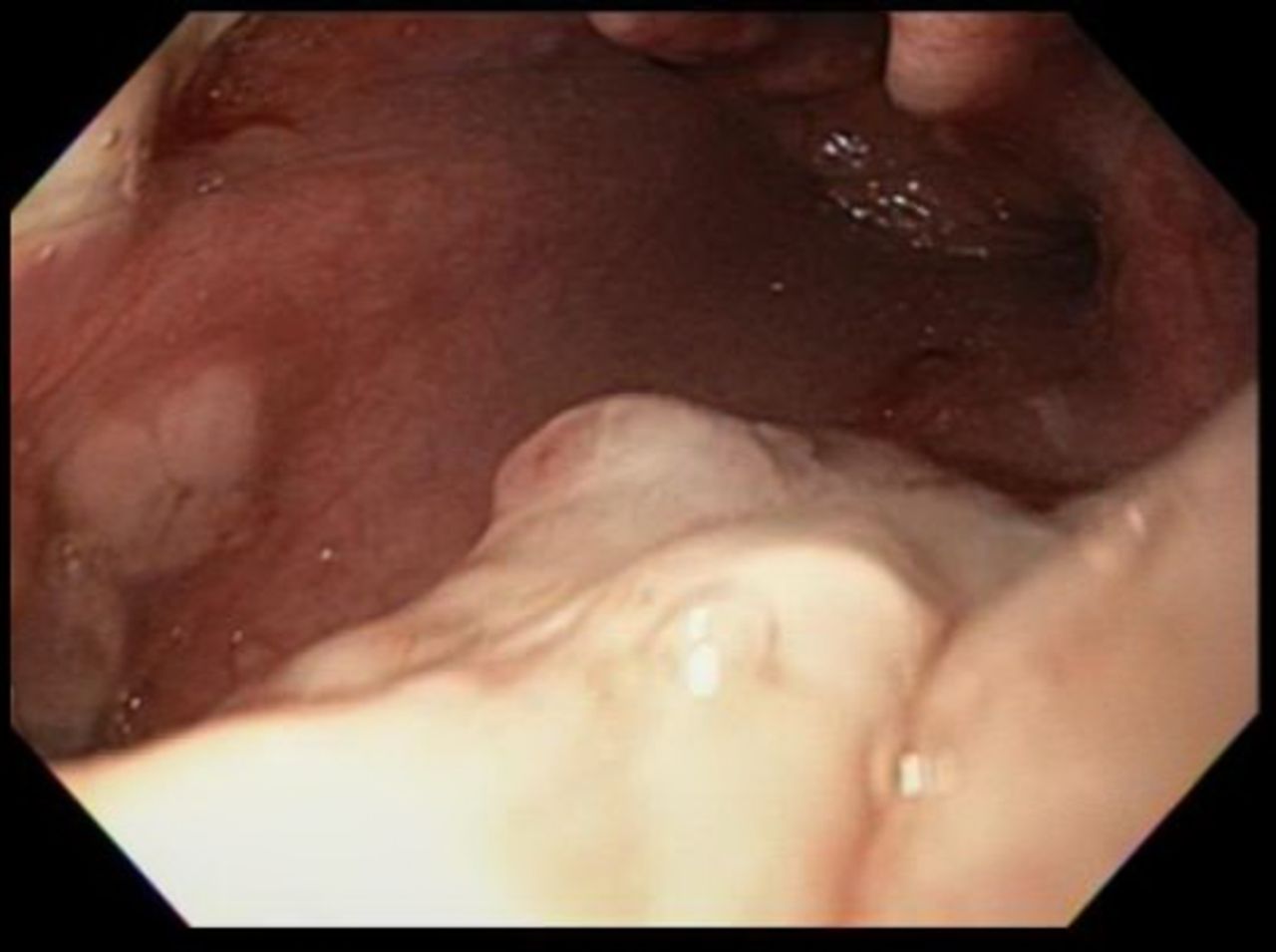
An upper gastrointestinal (GI) endoscopy was done on day 5 of hospitalisation showing whitish exudate and plaques at the base of her tongue, a 10 cm patch of whitish exudates in the oesophagus with underlying esophagitis (figures 2, 3). Esophageal brushings were obtained. Cytopathological examination of esophageal brushings showed cellular changes consistent with HSV infection. Viral culture and immunofluorescence staining from esophageal biopsy came positive for HSV-2 and negative for HSV-1, CMV and adenovirus.
Whitish exudate and plaques at the base of the tongue.

Patch of whitish exudates in the oesophagus with underlying esophagitis.
Intravenous acyclovir was started for biopsy-proven HSV-2 esophagitis. Immunosuppression was also lowered by halving the dose of mycophenolate mofetil. Adjustments in her tacrolimus dose were made due to interaction with fluconazole. During the treatment period her trough tacrolimus levels ranged between 5.5–8.4 ng/mL.
Outcome and follow-up
The patient’s symptoms improved with intravenous acyclovir. She was discharged home after 4 days of intravenous acyclovir, and was on oral valacyclovir as well as oral fluconazole until completion of 21 days of therapy. The patient followed up in the outpatient clinic 3 weeks later and reported complete resolution of her symptoms. She had a repeat esophagogastroduodenoscopy (EGD) which showed complete resolution of esophagitis (figure 4).

Esophagogastroduodenoscopy 3 weeks later showed complete resolution of esophagitis.
Discussion
Opportunistic infections in immunocompromised renal transplant patients are a fearful complication. The incidence is high in the first 6 months due to intense immunosuppression and transplant centres give prophylaxis against common infections during this period. Infectious esophagitis is among such opportunistic infections. Infectious esophagitis can present with symptoms including discomfort and difficulty in swallowing, pain while swallowing and retrosternal chest pain. Patients may have a prodrome of fever, malaise and nausea. The symptoms based on a previous reviews include dysphagia for both solids and liquids (37.5%), odynophagia (60.7%), chest pain (46.4%) heartburn, and/or vomiting.1
Candida is the most common cause of fungal infection of oral mucosa and oesophagus in immunosuppressed patients. Candida albicans or candida tropicalis are the commonly involved species.2 50%–75% of patients with esophageal candidiasis have oropharyngeal candidiasis. Oropharyngeal candidiasis most commonly presents in a pseudomembranous form with white plaques on the oropharynx, tongue, palate and buccal mucosa. The presence of oral thrush may be helpful in the diagnosis of fungal esophagitis;3 however, the absence of oral involvement does not exclude esophageal candidiasis. Diagnosis is usually made using endoscopy. Endoscopic evidence of candida lesions may include superficial erosions, ulcers and white plaques that can be severe, resulting in necrosis and perforation. Histopathological examination of biopsies shows the presence of yeast and pseudohyphae with invasion of mucosal cells and the culture of the biopsy specimen grows candida.4 5 The treatment includes a course of oral fluconazole. Itraconazole can also be used; however, it has more drug interactions than fluconazole. Intravenous antifungal therapy may be required in patients with severe disease and in those who cannot take oral medications. Other azoles, such as posaconazole and voriconazole are preferred in refractory cases. Interaction of azole antifungals with calcineurin inhibitors should be kept in mind while treating renal transplant patients with azoles.5 Echinocandins and amphotericin B can be used for azole refractive cases. If symptoms do not improve within 3 to 4 days, co-infection by a different agent should be strongly suspected.
On the other hand, HSV is second only to candida among pathogens that cause infectious esophagitis. HSV causes many types of GI pathologies ranging from mild, ulcer-like mucocutaneous lesions (herpes labialis, oropharyngeal ulcers) to esophagitis, hepatitis and colitis.6 Opportunistic infections commonly present in the first 6 months after transplantation and the reactivation of HSV usually occurs in the first 6 weeks. One study reported HSV-associated esophagitis in 5 of 221 renal transplant patients over an 8-year period. All these cases were associated with the treatment of acute rejection with high-dose steroids and antilymphocyte preparations.6 In contrast, our case is unique in presenting as a rare dual pathogen esophagitis in an immunocompromised renal transplant recipient 9 months after transplantation while she was on maintenance immunosuppression without any recent exposure to high-dose steroids or T-cell depleting agents.
In Herpes esophagitis, usually HSV-1 is the causative agent but HSV-2 esophagitis, though rare, has also been reported. HSV-2 usually causes genital herpes; rarely does it cause meningitis, pulmonary infections, esophagitis and proctitis. HSV infection is usually a reactivation of latent virus and can present in the early post-transplant period. It can spread from oropharyngeal lesions to the oesophagus. Diagnosis is usually made by endoscopy findings and histopathological examination. Endoscopic evidence of HSV esophagitis may reveal shallow, well-circumscribed ulcers with normal intervening mucosa, sometimes described as being ‘punched out’ or ‘volcano-like’. Coalescent ulcers with diffuse erosive esophagitis or pseudomembranous lesions may also be seen. Histological findings include multinucleated giant cells with ground-glass nuclei and eosinophilic inclusions (Cowdry type A inclusion bodies).4 5 Viral culture and immunofluorescence testing aid in the diagnosis. HSV DNA PCR of the biopsy sample is the most sensitive test for the detection of HSV infection with a rapid turnaround time. The treatment of HSV esophagitis in an immunocompromised patient involves a course of oral acyclovir. Patients with severe disease require a longer course or intravenous acyclovir treatment. Patients with difficulty in swallowing usually require hospitalisation for intravenous acyclovir and can be switched to oral therapy when clinical improvement is evident.5 Intravenous hydration and close monitoring of renal function is advised during treatment with intravenous acyclovir due to the possibility of crystals associated acute kidney injury. Oral famciclovir and valacyclovir allow for a decreased pill burden though at a higher cost of treatment. Acyclovir-resistant cases may be encountered in immunosuppressed patients and may need treatment with foscarnet.
In CMV infection of the GI tract, colon and oesophagus are the most common sites involved. Symptoms of CMV esophagitis may include difficult or painful swallowing and epigastric pain.7 EGD is the imaging study of choice. Large shallow ulcers in the distal oesophagus and at the lower esophageal sphincter are a common finding but diffuse esophagitis can also occur. Ulcers tend to be longitudinal or linear.5 The tissue sample is usually sent for histopathological examination, antigen detection, viral culture and PCR. On histopathological examination, mucosal inflammation, along with cytomegalic cells that have large nuclei and intranuclear or intracytoplasmic inclusions classically described as ‘owl’s eye appearance’, are seen. Intravenous ganciclovir is usually used for the initial treatment of the CMV disease. Oral valganciclovir is also considered an effective drug due to its high bioavailability.8 Foscarnet is often used for gancyclovir-resistant CMV disease,9 either alone or in combination with gancyclovir. Cidofovir can also be used; caution is advised due to the nephrotoxic potential of these medications. Genotype resistance testing and expert consult are advised in such cases.10
Candida and CMV may coexist in up to 20% of the patients; however, simultaneous infection by Candida and HSV is even less common. A prospective study reported 100 immunocompromised patients with AIDS and esophagitis, 33 of whom had candida alone, 22 patients had coexistent candida and CMV while two patients had candida along with CMV and HSV.11 The potential mechanism for co-infection might be from initial injury to the esophageal epithelium by HSV and disruption of the mucosal barrier, which provides a supportive environment for candida, a normal commensal of the oral cavity.12 In our literature review, we were able to find a few case reports of concomitant infection of these pathogens in immunocompetent hosts.13–15 Kumar et al, describe a 57-year-old man with esophageal tuberculosis with coexisting HSV and candida, 8 months after renal allograft transplant.16 Cases of dual infection of the oesophagus by candida and HSV-2 in immunocompromised renal transplant recipients are rare.
Our patient presented with common symptoms of fever, dysphagia and odynophagia along with oropharyngeal thrush and whitish tonsillar exudates. Candida was suspected, prompting us to initiate fluconazole for antifungal therapy. She did not have any mucocutaneous ulcers or vesicles suggestive of HSV. While the presence of oral thrush or herpetic vesicles may aid in diagnosis, a confirmatory diagnosis necessitates endoscopy with histopathological and microbiological examination. In a series of renal transplant recipients at a single institution, 97 patients with esophagitis underwent a biopsy during endoscopy. Esophageal candidiasis, CMV, and HSV were found in 33, 8 and five patients, respectively.17
Other causes of esophagitis including gastro-oesophageal reflux disease, medication-induced esophagitis, eosinophilic esophagitis, idiopathic aphthous ulcers of the oesophagus and radiation esophagitis should be considered in the appropriate clinical settings.
Learning points
Infectious esophagitis can be seen in patients after renal transplantation due to immunosuppressive therapy.
Dual infection is uncommon and co-infection can be missed on initial presentation leading to clinical complications.
A high level of suspicion is warranted in such high-risk patients.
Delayed or under-treatment can lead to worse clinical outcomes.
While treating esophagitis in immunosuppressed patients, empiric antifungal and as well as antiviral coverage should be considered in the appropriate clinical setting if the suspicion of a dual infection is high, before the final diagnosis is made.
Footnotes
Contributors IG: prepared most of the manuscript, reviewed the literature, was involved in patient care and management, consented the patient and provided the images. VK: wrote part of the manuscript, reviewed the literature. MS: wrote part of the manuscript. RK: reviewed, proofread the manuscript and provided feedback.
Funding The authors have not declared a specific grant for this research from any funding agency in the public, commercial or not-for-profit sectors.
Competing interests None declared.
Provenance and peer review Not commissioned; externally peer reviewed.
Patient consent for publication Obtained.