Article Text

Statistics from Altmetric.com
Description
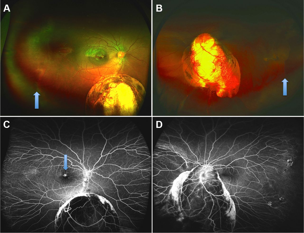
A 10-year-old boy presented with diminished vision in his right eye noted for 1 month. There was no history of prematurity. Family and systemic history was unremarkable. Best-corrected visual acuity (BCVA) was 20/120 in the right eye with +6 dioptres sphere and 20/40 in the left eye with +2 dioptres cylinder at 180°. Anterior segments were normal apart from the presence of inferonasal iris coloboma in both the eyes. The dilated fundus examination showed type III choroidal coloboma (Ida-Mann classification) in the right eye and type II choroidal coloboma in the left eye (figure 1A,B). Both eyes also had peripheral avascular retina separated by demarcation lines (arrows, figure 1A,B). Ultra-wide field fluorescein angiography (figure 1C,D) of both eyes revealed straightening of vascular arcades, supernumerary vascular branching and temporal avascular retina in both the eyes. A closer look at the macula revealed subfoveal choroidal neovascularisation (CNV, figure 2A), which was confirmed on swept source optical coherence tomography (figure 2A,B) and fluorescein angiography (figure 1C, arrow). A diagnosis of both eye choroidal colobomata with familial exudative vitreoretinopathy (FEVR) with right eye CNV was made and intravitreal bevacizumab was advised in the right eye.

UWF pseudo-colour photograph of the right (A) and the left eye (B) showing type-3 and type-2 choroidal coloboma respectively. The blue arrow marks line separating avascular peripheral retina from vascular retina. UWF fluorescein angiography (C,D) revealed straightening of vascular arcades, supernumerary vascular branching and temporal avascular retina consistent with familial exudative vitreoretinopathy. Arrow in (C) shows choroidal neovascularisation.

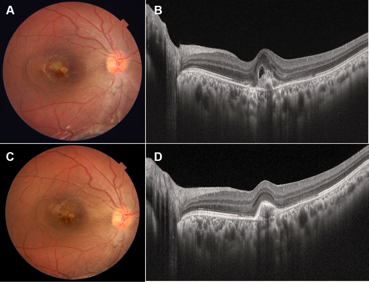
Colour fundus photograph (A) and swept source optical coherence tomography (SS-OCT) (B) show choroidal neovascularisation at the macula of the right eye. At 1 month after intravitreal bevacizumab injection, fundus photograph and SS-OCT (C,D) show resolution of intraretinal and subretinal fluid.
At 1 month after intravitreal bevacizumab, the CNV had regressed (figure 2C,D) and BCVA improved to 20/60. The parents declined further injections and was advised regular follow-up. The visual acuity was maintained at 6 months follow up with no signs of recurrence.
Familial exudative vitreoretinopathy is a bilateral asymmetric ocular hereditary disorder. It is characterised by peripheral avascular retina, neovascularisation and retinal detachment. About 50% of the cases can be linked to four causative genes FZD4, NDP, LRP5, and TSPAN12 which affect ‘wnt’ signalling pathway.1 Choroidal coloboma on the other hand occurs as a result of the failure of the embryonic fissure to close2 which can occur due to various genetic defects and environmental factors. The coloboma gene network developed from the genes associated ocular coloboma has sonic hedgehog and paired box 6 genes in the pivotal role.3 However, disruption of ‘wnt’ signalling pathway could also cause coloboma.4 An interaction of these genes may explain the coexistence of the FEVR with coloboma. CNV is a known rare complication associated with choroidal coloboma.5 This case highlights the occurrence of these two anomalies in a single patient which, to the best of our knowledge, has not been reported before.
Ultra-wide field angiography is useful for imaging of the peripheral retina, and it is particularly useful for providing panoramic images of the retina with a single click. This is especially relevant in paediatric patients6 or patients with poor vision, where creating a montage image may be time-consuming and a source of discomfort to the patient.
Learning points
Ultra-wide field angiography is useful for imaging of the peripheral retina, and it is particularly useful for providing panoramic images of the retina with a single click.
Familial exudative vitreoretinopathy may also be associated with coloboma of the fundus.
Footnotes
Contributors VK, SKP: acquisition of data, concept, writing manuscript, review.
Funding The authors have not declared a specific grant for this research from any funding agency in the public, commercial or not-for-profit sectors.
Competing interests None declared.
Provenance and peer review Not commissioned; externally peer reviewed.
Patient consent for publication Obtained.