Article Text

Statistics from Altmetric.com
Description
A healthy 31-year-old woman was referred to our department due to a hyperpigmented lesion, similar to a bruise, with a 6-year history and progressive growth, that has never disappeared. There was no trauma history. Physical examination revealed a violaceous plaque, with 10 mm of diameter, located on the left shoulder (figure 1). Dermoscopy revealed a violaceous background in conjunction with blue-whitish veil lesions without a peripheral pigment network, and fine linear vessels, equally distributed all over the surface of the lesion (figure 2). An incisional biopsy was performed and histological examination revealed a neoplasia composed of spindle cells arranged in a storiform pattern, some of them pigmented. Immunohistochemical study showed intense and diffuse expression of CD34 by neoplastic cells (figure 3C) and S100 protein by pigmented spindle cells (figure 3D). These results were compatible with a pigmented dermatofibrosarcoma protuberans (figure 3A,B). The patient underwent wide surgical excision. Thoraco-abdomino-pelvic CT scan was unremarkable. Currently, the patient is under regular follow-up, without any recurrence at 2 years.
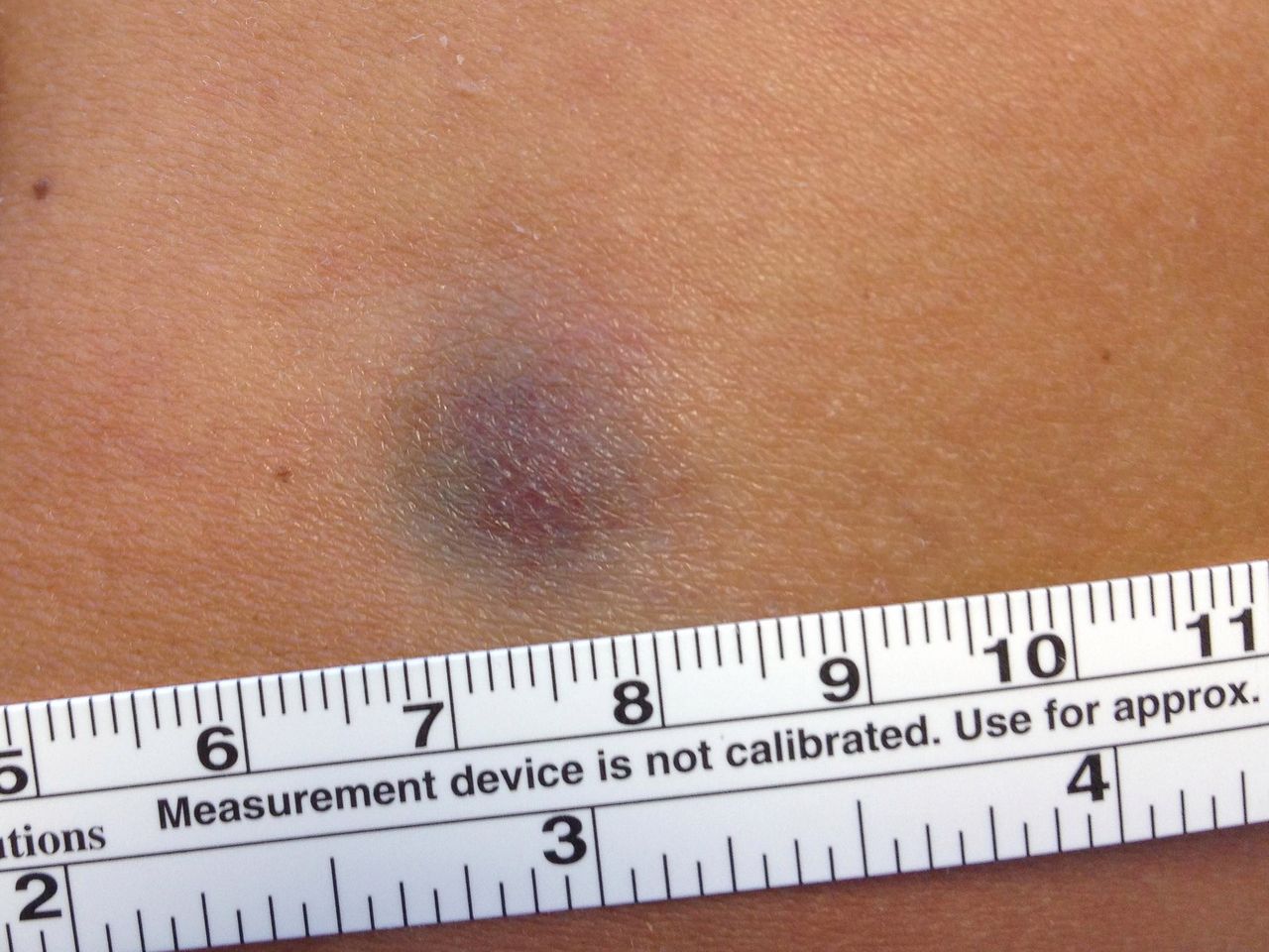
Bednar tumour located on the left shoulder.

Dermoscopy of Bednar tumour.

Microphotograph showing diffuse infiltration of dermis (A, H&E ×40) and subcutis (B, H&E ×100) by neoplasia composed of spindle cells arranged in a storiform pattern. Some spindle cells are pigmented. Neoplastic cells are immunopositive for CD34 (C, ×100) and pigmented spindle cells show expression of S100 protein (D, ×400).
Dermatofibrosarcoma protuberans (DFSP) is a rare cutaneous soft tissue sarcoma of intermediate malignancy, whose incidence peaks between the fourth and fifth decades of life. Bednar tumour, also known as pigmented DFSP, is a rare variant, accounting for less than 5% of all DFSP cases.1 It has a predisposition to affect the trunk, especially the back and shoulders, as seen in our case. Although it has significant subclinical extension and great capacity for local destruction and relapse, overall prognosis is favourable with prolonged survival and low rate of metastasis.2
The diagnosis of this tumour is difficult, as it can be mistaken, both clinically or histologically, for other skin tumours, including dermatofibroma, cellular blue nevus, fibrosarcoma and malignant melanoma.3 Therefore, any lesion with a prolonged evolution or that does not resolve spontaneously, even if clinically unsuspected, should always be biopsied to exclude malignancy.
We reported this case because of its rarity and unusual presentation and to highlight the importance of histological examination with immunohistochemical study to warrant a prompt and correct diagnosis and to provide an adequate management of the patient.
Learning points
Bednar tumour is an uncommon pigmented subtype of dermatofibrosarcoma protuberans.
It usually affects the trunk of young to middle-aged adults.
As clinical presentation may be heterogeneous, it is important to be aware of this entity and to perform histopathological analysis to make an early diagnosis.
Footnotes
Patient consent for publication Obtained.
Contributors The authors FTA, TP and CB contributed to the planning, conduction and report of the work. The authors FTA, SDC and TP contributed to the conception and design of the work. The authors FTA, SDC and TP contributed to the acquisition of analysis and interpretation of the results.
Funding The authors have not declared a specific grant for this research from any funding agency in the public, commercial or not-for-profit sectors.
Competing interests None declared.
Provenance and peer review Not commissioned; externally peer reviewed.