Article Text

Statistics from Altmetric.com
Description
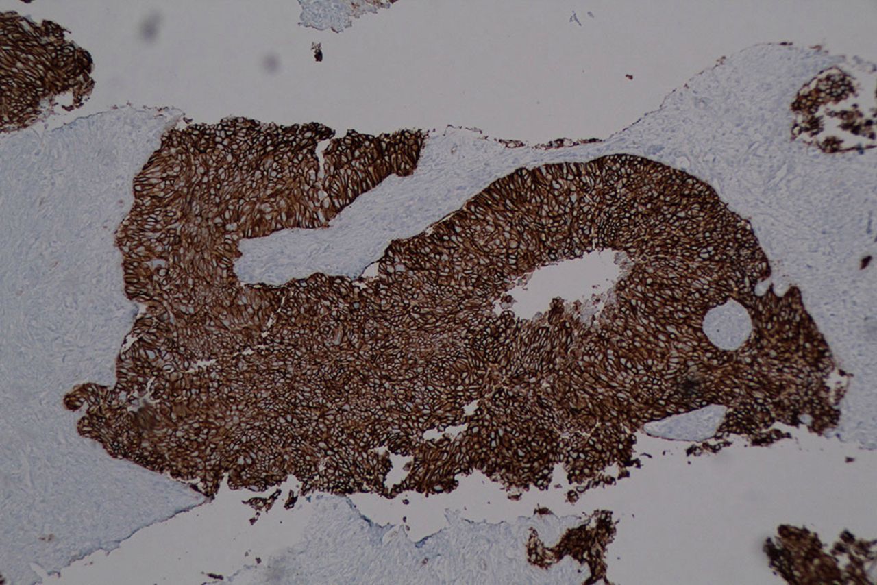
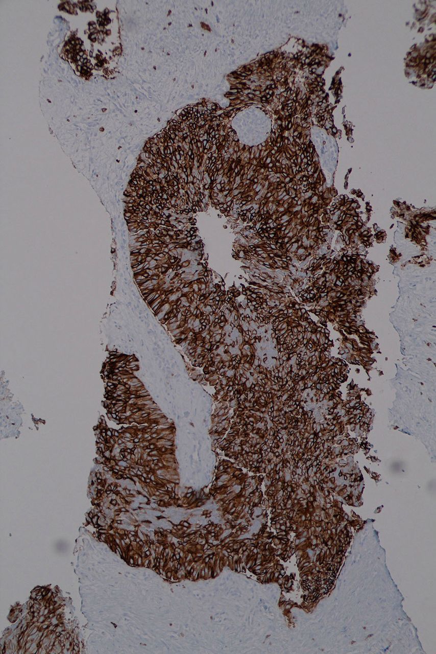
Primary thymic carcinoma is a rare malignancy and particularly basaloid type is an even rare subtype, with less than 20 cases published as individual case reports in the English literature.1–5 A 61-year-old female patient presented after falling, with concern for rib fracture. CT of abdomen/pelvis with contrast to evaluate for splenic injury revealed two left hepatic lobe masses with the largest measuring 5.1 cm concerning for metastatic disease. Positron emission tomography (PET) showed anterior mediastinal mass measuring 6.5×3.8 cm with standardized uptake value (SUV) 8.9 and two enlarged hypermetabolic liver masses. Ultrasound-guided liver biopsy showed a pleomorphic cell population not morphologically consistent with the liver primary (figure 1). Immunohistochemical stains were diffusely positive for pan-cytokeratin, GATA3, CD117, P63, CD5 and showed scattered positive cells for CK7. The neoplasm was negative for CK 20, SatB2, ER, PR, GCDFP-15, mammaglobin, TTF-1, Hep Par 1, synaptophysin, chromogranin and CK 5/6. Coexpression of CD 117 and CD5 is typical of thymic carcinoma (figures 2 and 3). The histological features and the immunohistochemical staining pattern were consistent with metastatic basaloid-type thymic carcinoma. The patient was subsequently started on carboplatin area under the curve (AUC) 5 and Taxol 200 mg/m2. The patient is currently status post 11 cycles of chemotherapy and interim scans showed response to treatment. Thymic carcinomas are rare aggressive tumours with poorer prognosis and higher mortality than thymomas as they often metastasize to regional lymph nodes and extrathoracic sites.6 Given the rarity of this malignancy, chemotherapy options are extrapolated from phase II studies. Carboplatin/paclitaxel is the preferred first-line therapy with an overall response rate, 22%–36%.7 Further long-term follow-up and examination of additional cases are needed to understand exact pathophysiology and assess response to additional chemotherapy.

Liver biopsy showing pleomorphic cell population.

Immunohistochemical stain highlighting diffuse positivity for CD5.

Immunohistochemical stain highlighting diffuse positivity for CD117.
Learning points
Basaloid type of thymic carcinoma is a rare malignancy with less than 20 cases published in the literature.
Coexpression of CD117 and CD5 on immunohistochemistry is typical of thymic carcinoma.
Thymic carcinomas do have a poorer prognosis and higher mortality than thymomas as they often metastasize to regional lymph nodes and extrathoracic sites.
Footnotes
Contributors SM and HHR worked on the abstract, PNC provided with a pathological description along with images, and KC revised and edited the abstract.
Funding The authors have not declared a specific grant for this research from any funding agency in the public, commercial or not-for-profit sectors.
Competing interests None declared.
Patient consent for publication Obtained.
Provenance and peer review Not commissioned; externally peer reviewed.