Article Text

Statistics from Altmetric.com
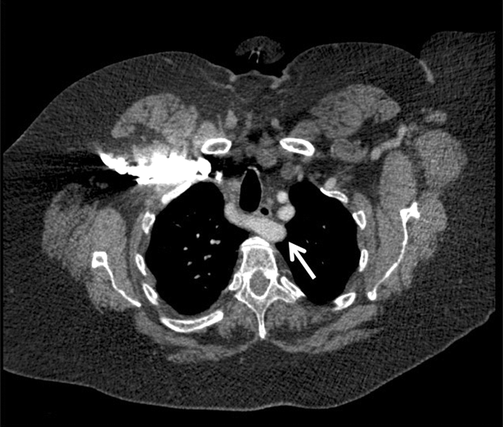
A 54-year-old woman was evaluated for 6 months history of solid food getting stuck to her oesophagus behind the upper half of sternum. On examination, a difference of 14 mm Hg was noted between the systolic blood pressures of her arms, the left being higher. A review of her admission day chest computed tomography (CT) scan that was obtained for a suspected pulmonary embolism (patient initially presented with shortness of breath related to her obstructive pulmonary disease) was undertaken. The CT scan revealed an aberrant right subclavian artery (ARSA) coursing from left to right posterior to the oesophagus (fig 1). An oesophageal endoscopy showed a transverse luminal narrowing with functional obstruction 22 cm from the incisors (fig 2). Arterial pulsations were noted in the posterior wall at the site of narrowing. An endoscopic ultrasound confirmed the subclavian artery (fig 3). A diagnosis of dysphagia lusoria due to ARSA was entertained. Given the patient’s mild degree of dysphagia and an abundance of significant comorbidities, a surgical treatment was deferred and the patient was discharged with dietary advice.

Computed tomography scan of the patient’s chest at the level of third thoracic vertebra shows an aberrant right subclavian artery coursing from left to right posterior to the oesophagus (arrow).

Oesophageal endoscopy shows normal looking mucosa and transverse narrowing at 22 cm from incisors.

Endoscopic ultrasound (radial view, colour mode Doppler) showing an artery (arrow) posteriorly coursing and abutting onto the oesophagus.
ARSA is the most common embryological abnormality of aortic arch and is seen in ∼1% of the population.1 The ARSA arises from the aorta distal to the origin of the left subclavian artery and follows an encircling retro-oesophageal course between the oesophagus and the spine. In its course it can cause extrinsic compression of the oesophagus leading to dysphagia, yet most are asymptomatic. It was first described in 1794 by David Bayford as “lusus naturae” where lusus is Latin for “sport” or “freak” of nature.2 In a retrospective review of 7513 unselected oesophageal ultrasound examinations, 27(0.36%) were found to have ARSA.3 In the same series, 10 out 14 patients had their anomalies missed on their CT chest films that was later detected on CT re-review after ARSA documentation on oesophageal ultrasound. Most cases with dysphagia respond to dietary modification, but surgical repair (transection and reimplantation of the artery) may be undertaken in selected cases.
This vignette emphasises the potential role of careful CT reading in suspected ARSA cases.
Footnotes
Competing interests: none.
Patient consent: Patient/guardian consent was obtained for publication