Article Text

Statistics from Altmetric.com
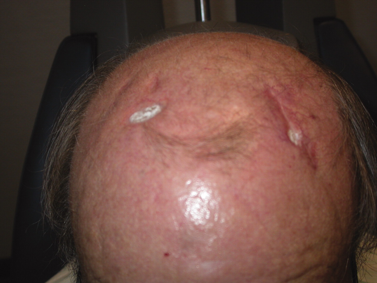
A 77-year-old man presented to his general practitioner with a painful scalp rash (fig 1). This worsened over 9 weeks and he was referred to a dermatologist. The dermatologist presumed the lesions were healing vesicles from herpes zoster and prescribed topical acyclovir. One week later he presented to the ophthalmology department following a 3 day history of deteriorating left vision. He also noted symptoms of lethargy and jaw claudication over the previous 2 weeks. On examination visual acuity was 6/12 OD and counting fingers OS. Erythrocyte sedimentation rate (ESR) and C reactive protein (CRP) were raised and temporal artery biopsy confirmed giant cell arteritis (GCA). Intravenous methylprednisolone 1 g stat and oral prednisolone 80 mg once daily were instituted. Since commencing his treatment his inflammatory markers improved as did his scalp lesions (fig 2). The vision in his left eye, however, has remained at counting fingers.


The main distinguishing factor from herpes zoster in this case is that the lesions were bilateral. If they had been correctly recognised as signs of GCA, prompt treatment may have saved the left vision.
Common presenting symptoms of GCA include headache, jaw claudication, scalp tenderness, and polymyalgia rheumatica. Scalp necrosis (SN) is a rare but known presentation.1,2 Currey3 reviewed 24 cases of GCA that presented with SN. Of these, 13 were referred to a dermatologist and three were misdiagnosed as herpes zoster. The review showed a higher incidence of irreversible visual loss and mortality which may reflect a more aggressive form of vasculitis.
Footnotes
Competing interests: None.
Patient consent: Patient/guardian consent was obtained for publication