Article Text

Statistics from Altmetric.com
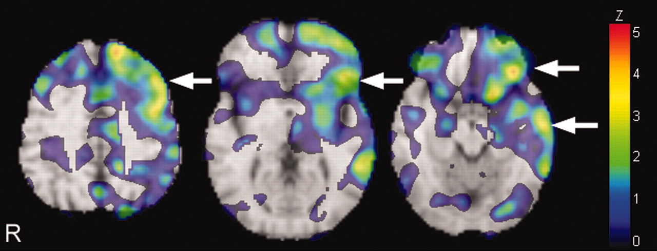
A 62-year-old right handed woman experienced a sudden decrease in spontaneity and a concurrent deterioration of memory. Her consciousness was clear. Some points, such as recalling the names of three simple preregistered objects after 5 mins and calculation, were deducted from the Mini-Mental State Examination (MMSE) score (23/30). She was oriented to time and place. The Wechsler Memory Scale-Revised showed verbal rather than spatial memory impairment (the spatial memory index/verbal memory index difference score was 26, a significant value). No abnormalities were observed in any other neurological findings. Her electroencephalogram was normal. Magnetic resonance imaging (MRI) showed a regional lacunar infarction in the thalamus (fig 1). MR angiography showed stenosis of the left posterior cerebral artery, but there was no sign of obstruction or stenosis of the internal carotid arteries. Her single photon emissions computed tomography (SPECT) scan Z score decreasing image showed decreased blood flow in the frontal, temporal, and parietal lobes (fig 2). Her memory improved at 6 months from onset (MMSE score 29/30).

Axial T1 weighted (A) and T2 weighted (B) images of the brain magnetic resonance imaging (MRI) demonstrate a lacunar infarction in the region encompassing the anterior nucleus of the left thalamus, the anterior ventral nucleus, and the lateral ventral nucleus (arrow). The MRI was performed 6 months after the onset of symptoms.
An examination of the Z score decreasing image of N-isopropyl-p123I iodoamphetamine single photon emission computed tomography (123I-IMP SPECT) revealed hypoperfusion in the frontal lobe, temporal lobe, parietal lobe, caudate nucleus, and putamen (arrow). The Z scores were >2.0, which are significant values. The spatial distribution of abnormal cerebral blood flow (CBF) was evaluated using the Neurological Statistical Image Analysis Software,1 and the value of the Z scores were calculated. Relative to the control mean, a positive Z score represented a decrease in the patient’s regional CBF.
The patient had a thalamic infarction in the dominant left hemisphere. The SPECT images revealed extensive blood flow defects in the corresponding cerebral hemisphere where the afferent fibres from the thalamus terminate. She described recent memory disturbances with a substantial decline in verbal memory performance, which is consistent with the symptoms of thalamic amnesia. In view of these facts, it could be concluded that her amnesia resulted from thalamic lacunar infarction.2,3
It is therefore necessary to consider thalamic lacunar infarction as a potential underlying aetiology of amnesia.
Footnotes
Competing interests: none.
Patient consent: Patient/guardian consent was obtained for publication