Article Text

Statistics from Altmetric.com
Description
A man in his 70s was referred for an internal medicine consultation with complaints of dysphagia for solid foods and liquids for 3 months, severe weight loss (6 kg in 2 months) and dysphonia for about a year. He had already been evaluated and discharged by an otorhinolaryngologist. He had Parkinson’s disease as relevant medical history. Laboratory tests were within normal parameters. Upper digestive endoscopy showed an extrinsic compression in the upper 1/3 of the oesophagus. A CT scan of the cervical, thoracic, abdominal and pelvic regions was normal. Hyperostosis with osteophytic formation was especially apparent at C2 level (figure 1) in the cervical spine CT scan and in a 3D reconstruction (figure 2). A barium swallow study demonstrated an exuberant anterior osteophytosis from C3 to C7, with posterior oesophageal compression. Due to his severe symptoms, the patient was referred for an orthopaedic consultation for evaluation for possible surgical treatment with anterior cervical osteophyte resection.
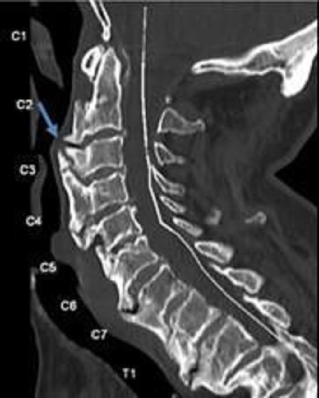
Cervical spine CT scan showing the compression of posterior oesophagus at C2 level, with an exuberant anterior osteophyte.

Cervical spine CT scan 3D reconstruction showing the compression of posterior oesophagus at C2 level, with an exuberant anterior osteophyte.
Foristier’s disease, also known as diffuse idiopathic skeletal hyperostosis, is a condition characterised by non-inflammatory ossification of the anterior vertebral longitudinal ligament with the development of osteophytes.1–3 It is mainly observed in men over 50 years of age,2 with a prevalence of 8%–10% over 65 years old.1 Its etiopathogenesis is not yet clear.1 2 The most commonly affected area in the cervical vertebrae is the lower part (C4–C7)2 and the involvement of the upper cervical vertebrae is very rare.1 Because of its location and proximity to the oesophagus, dysphagia can be seen in 0.6%–1% of patients with this condition.1 The diagnosis is radiological2 and the gold standard is the CT scan.1 In cases with severe symptoms such as progressive dysphagia or excessive weight loss, a surgical approach is recommended.1 4
The case represented involved an upper cervical vertebrae (C2) that was the cause of dysphagia by oesophageal compression, which is very rarely reported in literature.
Learning points
Although it is most commonly seen in otorhinolaryngology practice, diffuse idiopathic skeletal hyperostosis is a rare cause of dysphagia that should be considered in its differential diagnosis.
Even though the dysphagia can be explained by a neurological disease such as Parkinson’s disease, other conditions might need to be ruled out.
The correct diagnosis is crucial in order to initiate a multidisciplinary approach and give the proper treatment and adequate follow-up to prevent further complications.
Depending on the patient’s characteristics, the severity of the symptoms and any complications, it can be treated surgically or with a conservative approach.
Ethics statements
Patient consent for publication
Footnotes
Contributors CPR conceived the original idea, wrote the manuscript with support from CB, FV and LS. CB helped in writing the work. FV and LS helped to supervise the work.
Funding The authors have not declared a specific grant for this research from any funding agency in the public, commercial or not-for-profit sectors.
Case reports provide a valuable learning resource for the scientific community and can indicate areas of interest for future research. They should not be used in isolation to guide treatment choices or public health policy.
Competing interests None declared.
Provenance and peer review Not commissioned; externally peer reviewed.