Article Text

Statistics from Altmetric.com
Description
Fungal keratitis manifests clinically as epithelial ulcer, pseudopodia, corneal immune ring, hypopyon and endothelial plaque.1 It is an uncommon presentation that indicates severe infection with penetration of fungal elements into the anterior chamber.2
A man in his 30s visited the outpatient department following trauma to the right eye with sugarcane leaf 4 days ago while working on the farm. He presented with redness, watering, throbbing pain and blurred vision in the right eye. He did not have any comorbidity. The best corrected visual acuity was 6/18. On slit-lamp examination, we noted circumciliary congestion, 6×4 mm white stromal infiltration situated paracentrally extending to the periphery at six o’clock with feathery margins, and three satellite lesions peripheral to the lesion. It was associated with 1×2 mm endothelial plaque. There was no epithelial defect on fluorescein staining. The surrounding corneal stroma revealed mild keratitis (figures 1 and 2).

Slit-lamp microscopic image of the right eye taken at presentation showing 6×4 mm white stromal infiltrate (blue arrow) with feathery margin (red arrow) and satellite lesion (white arrow).
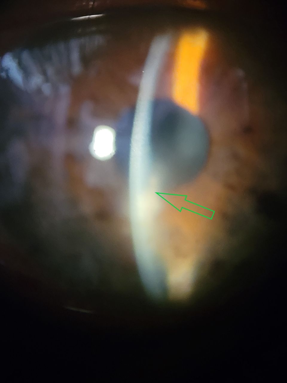
Slit-lamp microscopic image of the right eye taken at presentation showing endothelial plaque (green arrow).
Based on clinical evaluation and KOH (Potassium hydroxide) mount report, fungal keratitis with endothelial plaque was diagnosed. He was treated with oral Itraconazole 100 mg two times per day, topical Natamycin 0.5% eye drops, Voriconazole 1% eye drops and Homatropine 2% eye drops. The patient was followed up every day for 3 days and later for 1 week; during these follow-ups, clinical reduction in the size of infiltrate and stromal oedema (figure 3) attests to it. At 1-month follow-up, the patient was completely asymptomatic, with improvement in vision to 6/9 was noted with the presence of macular grade opacity measuring 2×3 mm paracentrally at six o’clock position (figure 4).

Image taken at 1-week follow-up showing a remarkable reduction in stromal infiltrate (blue arrow) and endothelial plaque (green arrow).

Image taken at 1-month follow-up showing 2×3 mm paracentral macular grade opacity (orange arrow).
Fungal corneal ulcers are expected to be prevalent more in areas where agriculture is the principal occupation among the population.3 Goel et al concluded that sugarcane injuries are more common among farmers.4 Krishnaiah et al, in their study, found that injury with vegetable matter such as a thorn, branch of a tree, plant secretion, etc (45.3%) was the primary cause of trauma reported in fungal keratitis.5 Qi et al found that endothelial plaques signify hyphae-infiltrating Descemet’s membrane, which is usually associated with poor response to medical treatment and worsening visual prognosis.6 Endothelial plaque is an uncommon presentation more specifically related to fungal keratitis, with 7.5% in a study by Upadhyay et al. It requires aggressive treatment and surgical intervention. It is associated with poor visual prognosis.6 7 The newer modalities in diagnosing endothelial plaques are anterior segment optical coherence tomography and in vivo confocal microscopy.8 In the present study, we identified endothelial plaque by slit-lamp examination. The aggressive topical medication helped early healing of keratitis with minimal residual opacity.
Hence, we conclude that this is an uncommon case of fungal keratitis with an endothelial plaque and no epithelial defect, which showed remarkable improvement without surgical intervention.
Patient’s perspective
Prior to being injured by a sugarcane leaf while working on the farm four days ago, my vision had been quite stable. However, after the injury, I experienced redness, wetness, pain, and blurred vision. This affected my day-to-day operations. I consulted the ophthalmologists, who informed me that I have an uncommon infection presentation that needs fast and rapid treatment. I adhered completely to the recommendations and followed up frequently. My vision had much improved during the follow-up appointment after a month, and I could now perform my everyday tasks with ease. I've been told to use safety glasses in order to avoid similar situations in the future.
Learning points
The early presentation of an endothelial plaque, which is a definite sign of fungal keratitis, can help ophthalmologists to suspect fungal keratitis.
The presentation of an endothelial plaque indicates the need for aggressive treatment to prevent vision-threatening complications of fungal keratitis.
Sugarcane leaf injury is commonly associated with fungal keratitis, and a detailed assessment of the patient can help in the early diagnosis and treatment.
Ethics statements
Patient consent for publication
Footnotes
Contributors RRM was involved in clinical evaluation, investigation and management. NAS was involved in literature search and drafting the manuscript.
Funding The authors have not declared a specific grant for this research from any funding agency in the public, commercial or not-for-profit sectors.
Case reports provide a valuable learning resource for the scientific community and can indicate areas of interest for future research. They should not be used in isolation to guide treatment choices or public health policy.
Competing interests None declared.
Provenance and peer review Not commissioned; externally peer reviewed.