Article Text

Abstract
A woman in her 70s suffered a motor vehicle accident, resulting in an intraarticular, distal femoral fracture with a unique fracture pattern. A 3D-printed model was obtained using the preoperative CT scan for surgical planning. Anatomic reduction was achieved using a medial parapatellar approach and the fracture was fixed with a medial buttress plate and interfragmentary screws. After 18 months, the patient presented a fair Knee Society Score of 62.
- Orthopaedic and trauma surgery
- Orthopaedics
This is an open access article distributed in accordance with the Creative Commons Attribution Non Commercial (CC BY-NC 4.0) license, which permits others to distribute, remix, adapt, build upon this work non-commercially, and license their derivative works on different terms, provided the original work is properly cited and the use is non-commercial. See: http://creativecommons.org/licenses/by-nc/4.0/.
Statistics from Altmetric.com
Background
Distal femoral fractures are infrequent injuries with a prevalence of 0.5% of all fractures and 3%–6% of adult femoral fractures.1 2 They have a bimodal distribution, affecting young individuals through high energy mechanisms and the elderly through low energy mechanisms. The mechanism of injury in most distal femoral fractures is thought to be axial loading with varus/valgus or rotational forces.3 Anatomically, the distal femur is formed by two condyles and a supracondylar region. The distal femur has a trapezoidal shape in axial view, with a lateral slope of 10° and a medial slope of 25°. In the coronal plane, the anatomic lateral distal femoral angle measures 81°–84°.4 The distal epiphysis of the femur develops at around 36 weeks of gestation and merges with the metaphysis at around 14–19 years.5 Epiphysiolysis of the distal femur occurs at paediatric age and corresponds to about 5% of all physical injuries, which can be complicated by growth arrest, limb length discrepancy and angular deformities.6
The most frequently used classification is the Arbeitsgemeinschaft Osteosynthese/Orthopaedic Trauma Association, with Neer and Seinsheimer classifications being of historical interest (table 1).7–13
A review of the classification systems for distal femoral fractures 8 10 11 13
An article published by Xie et al in 2017 mapped the fracture pattern of 75 Hoffa fractures by CT and concluded that Hoffa fractures are more frequent in the middle third of the lateral condyle, extending from an anteroinferior position for posterosuperior and from anterolateral for posteromedial. In order to analyse the fracture patterns, the authors defined two angles, the alpha angle was defined as the acute angle formed by the fracture line and the posterior condylar axis and the beta angle was defined as the acute angle formed by the fracture line and the posterior cortex of the distal femoral shaft.12
Unlike Hoffa fractures (table 2), reports of unusual distal femoral fracture patterns are scarce. Gahlot et al reported a case of a triplane fracture in a 30-year-old adult.14
A review of the clinical cases of Hoffa fractures in recent literature19–35
There are no standardised treatment guidelines for distal femoral fracture as, for example, with fractures of the proximal femur.15 Surgical treatment is indicated for most intrarticular and extrarticular fractures with deviation; however, osteosynthesis can be hampered by the presence of thin cortical bone, wide spinal canal, poor bone quality, displacement of fracture secondary to muscle pull and fracture comminution. To overcome these difficulties, there has been a significant advance in the methods of fixation and minimally invasive approaches, with the latest advances being locked anatomical plating, retrograde nailing and distal femoral replacement. Retrograde nails are mainly indicated in type A and in type C fractures with an intrarticular line capable of undergoing open reduction and fixation with interfragmentary screws. Lateral anatomical plates are mainly indicated for types A and C fractures. Distal femoral replacements are used in comminuted distal femoral fractures in low demand patients with poor bone stock. Most type B fractures can be treated with interfragmentary compression of the joint surface with screws and buttress plate.4 15 16 The main goals of surgical treatment of the distal femur fractures are: anatomical reduction of the articular surface; restoration of limb alignment and length and stable internal fixation that allows early mobilisation.17 18
In the following case, the authors will present an unusual, complex distal femoral fracture and its treatment.
Case presentation
A woman in her 70s, previously autonomous, was admitted to the emergency room after a motor vehicle accident with trauma to the lower limbs. She presented with oedema, pain and deformity of the right knee, without neurovascular deficits.
Investigations
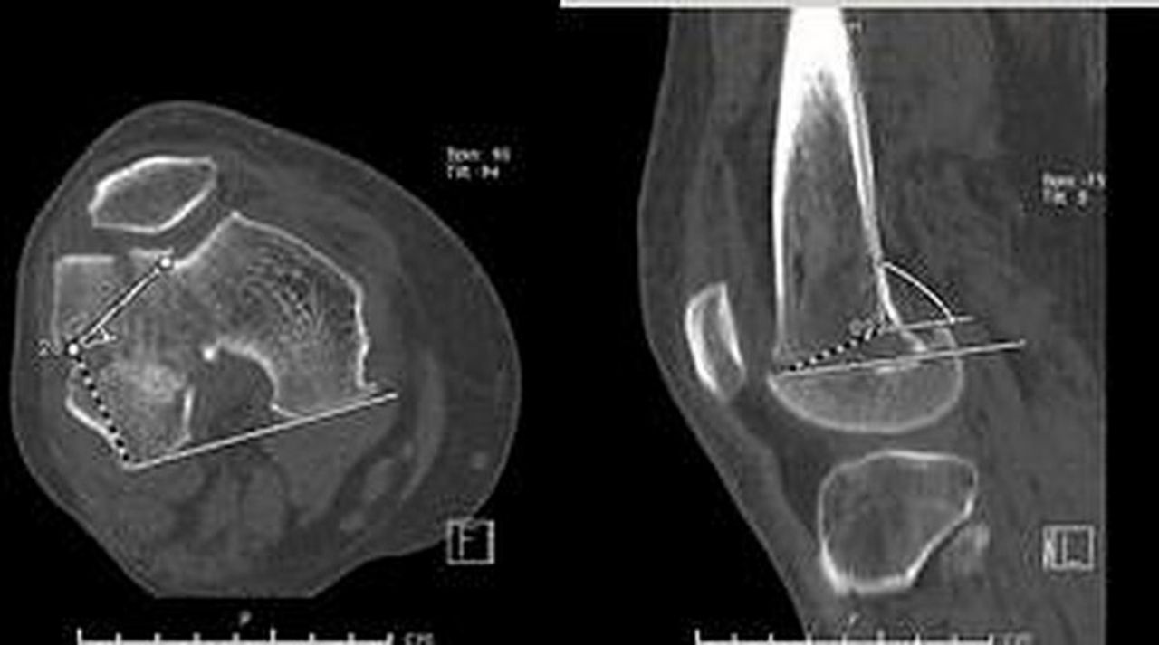
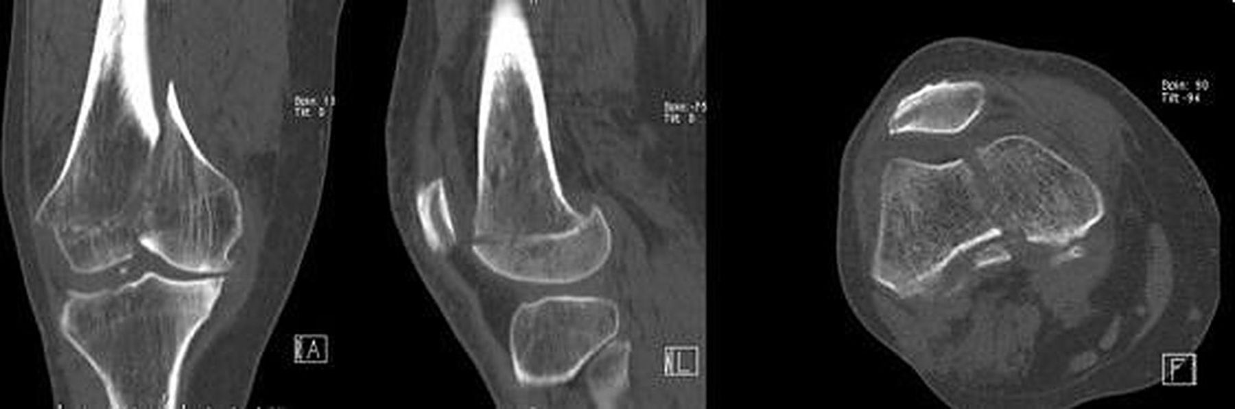
Radiological analysis revealed fracture of the right distal femur, with intrarticular extension (figure 1). A CT scan with 3D reconstruction was obtained to further define the fracture, which presented a medial fragment, of large dimensions, with a fracture line that extended from the intercondylar notch to the medial cortex and a lateral fragment with an horizontal-oblique fracture line from superior-medial to inferior-lateral, through the epiphysial scar (figures 2 and 3). A 3D model was obtained to aid in preoperative planning (figure 4).
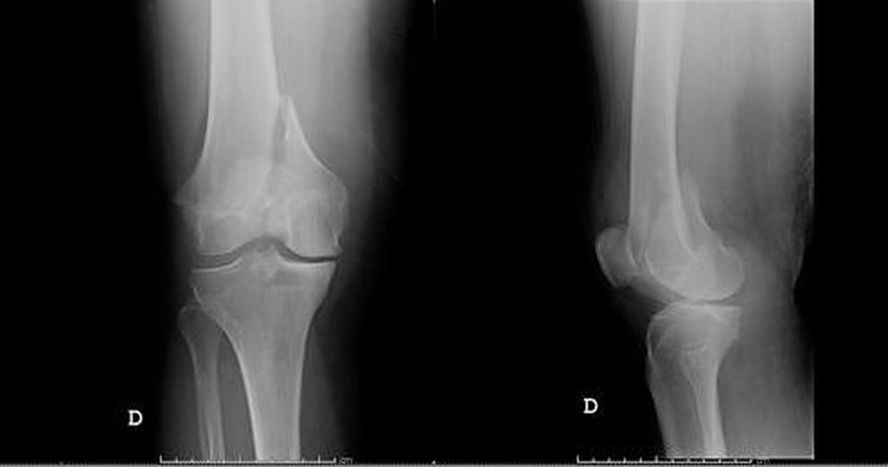
Preoperative radiography of the distal femur. Antero-posterior and lateral view.

Preoperative CT scan of the distal femur. Sagittal and axial view to the top. Left—coronal view. Middle—sagittal view at the level of the lateral fragment. Right—axial view at the level of the medial fracture fragment.

Reconstruction images to distal femoral fracture.

: 3D model of the distal femoral fracture obtained with 3D printing.
Differential diagnosis
After a knee trauma, a simple contusion, ligamentous injury or fracture may be present. X-ray analysis was sufficient for the diagnosis of a distal femur fracture although insufficient for the characterisation of the fracture and preoperative planning. Only after the CT scan was obtained, we were able to appreciate the full extent of the fracture.
Treatment
The lower right limb was temporarily stabilised with a cruropodalic plastered splint and the patient hospitalised.
On the 11th day after admission, the patient underwent surgical treatment. The patient was placed in the supine position, with a tourniquet on the thigh. A medial parapatellar approach was used. After temporary reduction with Kirshner wires, the lateral fragment was fixed using two interfragmentary screws, from inferior-medial to superior-lateral direction, perpendicular to the fracture line. Initially, the preoperative plan was the placement of indirect, cartilage saving screws in the lateral fragment, perpendicular to the fracture line but had to be revised to a direct position due to the lack of adequate distal bone stock observed during surgery. Due to the unexpected change of plans, headless compression screws were not available during surgery, as such, regular compression screws were used, with the heads buried beneath the cartilage to prevent secondary joint damage.
The medial fragment was reduced and compressed under direct and radiological visualisation using pointed reduction forceps and fixed with a buttress plate and standard screws (figure 5). Intraoperative and postoperative X-ray imaging was obtained, confirming the reduction (figure 6). The usage of CT scan would be more accurate for evaluating the construct, but it is not routinely used at our institution.

Intraoperative image of the osteosynthesis. Left—axial view of the interfragmentary lateral screws. Right—coronal view of the medial buttress plate.

Postoperative radiography of the distal femur. Antero-posterior and lateral view.
After surgery the patient started progressive rehabilitation with a mechanical splint and no weight bearing. Physiotherapy was suspended at 2 months post operatively due to COVID-19 infection.
Outcome and follow-up
After 18 months, the patient presented a fair Knee Society Score of 62. She presented occasional knee pain and tolerated weight bearing with a walking stick in the contralateral arm. The main limitation was due to a flexion contracture of about 20° (figure 7).

Range of motion of the right knee at 18 months follow-up. A flexion contracture of around 20° is present.
Discussion
There is no universally accepted classification for fractures of the distal femur, with the majority differentiating between extra-articular, intra-articular and isolated condylar fractures. These anatomical classifications fail to describe the conditions associated with fractures of the distal femur, namely the degree of deviation, comminution, degree of joint involvement, soft tissue damage, neurovascular structures, associated fractures, the degree of osteopenia and the presence of polytrauma, factors that determine the ‘personality’ of the fracture, which influence treatment and prognosis. The fracture pattern presented does not fit into any existing classification.
According to the AO criteria, a partial articular fracture, type B ‘involves part of the articular surface while the remainder of the joint remains intact and is solidly connected to the supporting metaphysis and diaphysis’ while in a complete articular fracture, type C ‘there is a disruption of the articular surface and the articular surface is completely separated from the diaphysis’. Based on the criteria presented, the fracture is classified as 33B2.3, a fragmentary, partial articular fracture, because of the anterior lateral articular surface segment that maintains continuity with the diaphysis (figure 3). However, given the fact that the articular surface contiguous with the diaphysis is small in size and does not contribute to the load distribution of the lower limb, it makes the fracture personality similar to a fully articular, multifragmentary fracture, 33C3.3, consisting of two large fragments: a medial fragment with an oblique fracture line, equivalent to an isolated fracture 33B2 and a lateral Hoffa-like fragment, in the axial plane similar to a Salter Harris type 3 fracture in paediatric patients.8
Despite the similarities, the lateral fragment cannot be considered a true Hoffa fracture because the line is in the axial and not in the coronal plane. Letournel’s classification defines oblique features as type III Hoffa fractures; however, it does not classify horizontal features.10
Recently, new classifications have been proposed for Hoffa’s fractures, based on CT images; however, these classifications also do not contemplate the existence of fractures in the horizontal plane.
According to Li et al classification, the lateral fragment is classified as I abc and although this classification is useful in assessing fracture comminution, it is not informative relative to the fracture configuration.11
According to Bagaria et al classification, the lateral fragment is classified as type I, according to size, but again, the classification is insufficient to describe the complexity of the fracture.13
Applying the Xie et al mapping technique, we obtained an alpha angle of −28° (the results described vary between −8.4 and 52.7) and a beta angle of 89° (the results described vary between −10.8 and 58.6) (figure 8).12 Comparing the measurements, we confirm that the presented fracture is roughly parallel to the axial plane, unlike Hoffa fractures that are perpendicular. The fracture also presents an obliquity that is the reverse to the ones seen in Hoffa fractures, that is, the fracture extends from inferior-lateral do superior-medial, unlike usual Hoffa fracture lines that extend from superior-lateral to inferior-medial, as seen in the coronal plane.
CT scan of the distal femur angle measurements. Left—alpha-angle of −28°. Right—beta-angle of 89°.
Of the clinical cases consulted, none had similar fracture traits as the one presented above.14 19–35
The treatment option chosen for this case is also debatable. In a regular Hoffa fracture, given the considerable sheer forces, it is subjected to during weight bearing, we would consider a three-screw construct, possibly with the addition of a buttress plate. The lateral condylar ‘Hoffa like’ fracture is horizontal in nature. Most of the forces applied during weight bearing are compressive, as such, a two-screw construct was considered sufficient. The small fragment size was also a concern regarding the placement of a third screw. Typical medial compression screws were not used to fix the medial fragment because of concerns regarding secondary fracture displacement with additional compression. A locking plate may have provided angular stability and a stiffer construct, but it is not mandatory for the buttress technique.
Some authors advocate the use of lateral precontoured locking plates regardless of the fracture line, arguing in favour of the mechanical advantage of the devices, that provides angular stability and the biological advantage of using less invasive stabilisation systems.16 However, this position is not consensual. Some studies indicate that the rigid construction of a locked plate impairs bone consolidation causing delay of consolidation and non-union.36 37
In our case, the authors opted to dismiss the lateral locking plate for the following reasons:
The lateral fracture line is distal, which would compromise the placement of the distal screws.
The position of the plate would interfere with the placement of the interfragmentary screws necessary for fixation of the lateral fragment to the metaphysis of the distal femur.
The fracture line is perpendicular to the plate, the use of a rigid osteosynthesis with locking plating could impair fracture consolidation.
The obtained osteosynthesis fulfilled the objectives of the surgical treatment. An anatomic reduction of the articular surface was achieved with stable internal fixation, which allowed early mobilisation of the limb and an acceptable functional result.16 Nevertheless, it has to be pointed out that certain treatment methods should be standard in the therapy of such fractures, namely, the usage of headless screws, intraoperative 3D imaging and postoperative CT scan to evaluate fracture reduction and implant placement.38 39
In conclusion, fractures of the distal femur can be complex and difficult to manage. The currently available classifications are insufficient to describe the complexity of the patterns found in clinical practice, and the surgical techniques currently used are unable to overcome all the problems associated with these fractures. More research is needed to evaluate the best treatment options and standardise patient care.
Patient’s perspective
I am pleased to be able to do my daily life with the help of my family and my walking cane.
Learning points
Distal femoral fractures may be a surgical challenge as the fracture patterns are diverse.
Parapatellar approach is a good alternative for complex intraarticular fractures as it allows good visualisation of the fracture and enables anatomic reduction.
3D modelling of the fracture pattern may aid in the preoperative planning.
Even after obtaining anatomic reduction, full recovery of the limb function may not be achieved.
The usage of headless screws, intraoperative 3D imaging and postoperative CT scan to evaluate fracture reduction and implant placement should be standard in the therapy of such fractures.
Ethics statements
Patient consent for publication
References
Footnotes
Contributors AC and PC were the main surgeons. JCR is responsible for the scientific review. AB is the corresponding author.
Funding The authors have not declared a specific grant for this research from any funding agency in the public, commercial or not-for-profit sectors.
Case reports provide a valuable learning resource for the scientific community and can indicate areas of interest for future research. They should not be used in isolation to guide treatment choices or public health policy.
Competing interests None declared.
Provenance and peer review Not commissioned; externally peer reviewed.