Article Text

Statistics from Altmetric.com
Description
A male patient, middle-age and BMI of 25.7 kg/m2 (weight 69 kg, height 1.64 m), was referred to the chronic pain unit 10 years ago for shoulder and residual thalamic pain following a cerebrovascular accident. This pain was treated with conventional analgesic therapy.
Five years ago, the patient reported a severe low back pain (LBP) with bilateral radiculopathy in the lower limbs without ‘red flags’. The medical history included diabetes, controlled hypertension and coronary artery bypass graft surgery. At that moment the patient was not taking any anticoagulation therapy. CT revealed moderate spinal stenosis in L3-L4 and root compression due to herniated disc protrusion in L4-L5.
Despite 6 months of several systemic multimodal analgesic therapies (increasing doses and rotation of strong opioids/adjuvants by the WHO ladder), the LBP prevented the patient from physiotherapy having a strong impact on daily living activities (DLA), mood and willingness to live.
The pain intensity (9-10 on the visual analogue scale) with bilateral radiculopathy (L4-L5 dermatomes distribution, Lasègue sign positive and without neurological deficits) had the characteristics of an intractable pain through conventional treatment. An invasive analgesic technique was then considered with patient’s consent. The patient was informed of the protocol safety when the criteria are followed strictly, and of the general and rare specific risks, namely dura-mater accidental puncture, subdural haematoma, urinary retention and local/systemic infection.
A protocol of continuous epidural technique with a tunnelled catheter (Box 1) was then initiated. A diagnostic and therapeutic technique was used with a single-shot lumbar epidural for immediate and prolonged relief of severe bilateral pain and functional disability (6.25 mg of levobupivacaine, 14 mg of betamethasone dipropionate and 1.5 mg of morphine). After 2 weeks without pain, there was an LBP recurrence requiring the protocol’s second stage to be followed (box 1).
Protocol of lumbar continuous epidural technique with tunnelled catheter
Patient inclusion criteria
Informed consent signed by the patient
Collaborative and oriented patient, able to acknowledge the procedure
Family support or caregivers able to understand the procedure
Transport availability for the patient with accompanying person
Absense of anticoagulation therapy and recent haemostasis evaluation
Absence of epidural technique contraindications
Pain unit with an available team to follow-up and execute sterile dressings
Available emergency service 24/7
First stage of the protocol
Diagnostic and therapeutic single-shot epidural bolus via interlaminar, foraminal or sacral approach performed at the moment of acute low back pain with unilateral or bilateral radiculopathy
Assessment of pain relief
10 mL bolus solution test administered with:
6.25 mg of levobupivacaine 0.625% (2.5 mL levobupi 0.25%), 14 mg of betamethasone dipropionate (or other non-particulate corticosteroid), 6.5 mL of saline solution (SS)
1-2 mg of morphine only in non-opioid-naive patients under previous systemic opioid medication
Second stage of the protocol
Performed in case of severe pain recurrence as long as the effectiveness of the single-shot epidural is confirmed
Undertake complementary analytical and imaging tests to further exclude contraindications to the technique
Continuous interlaminar or foraminal epidural technique is performed with or without imaging control at the lumbar intervertebral level
Subcutaneous double-needle tunnelling (figure 1) of the epidural catheter is performed in order to increase safety:
Promote safer tunnelling and better fixation of the catheter by reducing the possibility of its inadvertent externalisation
Reduce the risk and likelihood of infection by moving the catheter exit place away from the initial puncture
Reduce risk of contamination with extended periodical waterproof sterile dressings, always by the pain unit team, avoiding unnecessary exposure and manipulation by less-skilled professionals
An elastomeric infusion pump (EIP) with duration of 1, 2 or 5 days connected to the tunnelled epidural catheter is filled with:
Levobupivacaine doses between 10 and 16 mg/day (final concentration of levobupi 0.08%–0.125%) or ropivacaine 10–13 mg/day (0.08%–0.1%)
Morphine (2–5 mg/day) (optional in non-opioid-naive patients, if no contraindications)
Example: A 65 mL capacity EIP for 5 days and perfusion of 0.5 mL/hour, filled with 13 mL of levobupi 0.5% (65 mg) + 52 mL SS or 8.7 mL of ropivacaine 0.75% (65 mg) + 56.3 mL SS
Important: Before adapting the EIP, it has to be administered a 10 mL bolus of the same solution of the EIP via the epidural catheter (10 mL of levobupi 0.1% or ropivacaine 0.1%). Optionally, it can be associated to another corticosteroid dose and/or morphine
Analgesic doses are titrated according to patient characteristics, intensity of pain, complaints and conversion of systemic doses
Scheduling of serial EIP pick-up by the patient or caregiver
Final stage of the protocol
The preparation of the epidural catheter removal includes the use of substitutive therapeutic (specific dosages to each patient) in order to monitor the pain behaviour
Regular follow-up in the outpatient unit ensuring safety conditions and absence of complications
On removal, bacteriological analysis of the tip of the epidural catheter and blood culture are required
Assessment of the exclusion criteria for neurosurgical intervention
Systemic rescue analgesics are maintained for residual pain
Physiotherapy and rehabilitation until complete resumption of DLAs
Protocol of lumbar continuous epidural technique with double-needle tunnelled catheter elaborated by Carla Retroz-Marques and revised by authors

Epidural catheter tunnelling technique. (A) and (B) Double-needle technique for catheter protection; (C) tunnelling the catheter through the second needle ensuring an absence of catheter kinking; (D) catheter fixation and sterile waterproof dressing.
With the patient seated in his comfort position, a median interlaminar epidural approach was performed at L4-L5 intervertebral level, followed by 20G tunnelled catheter placement (figure 1) without complications. Simultaneously, with the gradual discontinuation of systemic opioids, the epidural perfusion analgesia was administered by sequential EIPs (capacity 65 mL for 5 days; 0.5 mL/hour; with 13 mg/day of levobupivacaine 0.1% + 5 mg/day of morphine), providing pain relief without adverse effects, while ensuring the restoration of DLA.
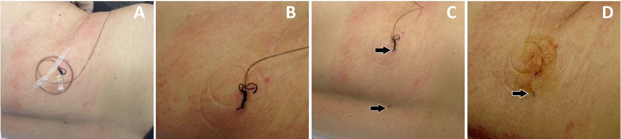
An MRI revealed a migrated hernia fragment lodged in the L4 root, without neurological compression. After 10 weeks, the epidural catheter was removed without complications nor inflammatory/infection signs (figure 2) (negative bacteriological analysis of catheter tip and blood culture).
Lumbar area after 10 weeks of the procedure. (A) Epidural tunnelled catheter fixed with surgical suture; (B) arrows pointing at the site of the initial puncture (completely healed) and at the emergence of the tunnelled catheter; (C) catheter exteriorisation; (D) intact catheter tip and skin orifice without infection or inflammatory signs.
After reassessment by the neurosurgical team, this favourable evolution of symptoms excluded criteria for surgical intervention.
During the last 4 years, the patient did not experience lumbosciatic pain recurrence and restored DLA. The original thalamic pain has been controlled with mild analgesic therapy.
This clinical case demonstrates that a technique widely used in other contexts can be adjusted for outpatient treatment of non-cancer pain, as long as the safety criteria are strictly respected. Studies confirm that continuous epidurals with opioids, anaesthetics and corticosteroids for LBP result in the control of disabling pain crises, reducing doses of systemic opioids with significant improvement of quality of life.1 2
Patient’s perspective
About 5 years ago, I had very intense low back pain, hindering my movements and dramatically affecting my daily life. Since various conventional treatment for more than 6 months failed to alleviate my pain, an epidural procedure was suggested, which I accepted having full trust on the pain unit team. During two and half months, I had a catheter ‘in my spine’ with a pump with medication that completely relieved my pain. This treatment also allowed me to recover my movements and I never felt again that very strong pain. For the last 4 years, I have had a normal life. Myself and my family are very grateful for the recommended treatment.
Learning points
Continuous outpatient epidural analgesia is a safe and effective procedure to alleviate disabling low back pain with uni/bilateral radiculopathy, if the protocol is strictly followed in carefully selected patients.
Despite being a widely used technique by specialists in chronic pain and obstetric anaesthesia, it is important to revisit it for low back pain since it is accessible and able to alleviate suffering due to refractory or intractable pain. However, the originality of the article is based on the specific context in which the protocoled technique is performed.
Despite the legitimate concern of healthcare professionals regarding the performance of invasive techniques in an outpatient setting, one should underline the capacity of this treatment to exponentially reduce the doses of systemic opioids and adjuvant analgesics, minimising side effects and risk of addiction while improving the patient’s quality of life.
Ethics statements
Patient consent for publication
Footnotes
Contributors CRM performed the therapeutic techniques and elaborated the protocol of the clinical case and participated in all stages, writing, reviewing and submitting the article. SSR and JA edited the article. AM did the final review of the article.
Funding The authors have not declared a specific grant for this research from any funding agency in the public, commercial or not-for-profit sectors.
Case reports provide a valuable learning resource for the scientific community and can indicate areas of interest for future research. They should not be used in isolation to guide treatment choices or public health policy.
Competing interests None declared.
Provenance and peer review Not commissioned; externally peer reviewed.