Article Text

Statistics from Altmetric.com
Description
The differential diagnosis of acute onset flank pain include renal colic, papillary necrosis, pyelonephritis and renal infarction from renal artery thrombosis or embolism. Here, we describe a case of acute onset flank pain caused by unilateral renal vein thrombosis (RVT) in a previously healthy patient. A 43-year-old woman was admitted with sudden onset of left-sided flank pain 72 hours prior to admission. There was no history of trauma, renal stone disease, dysuria, weight loss, oedema or family history of thrombotic episode. She had hypothyroidism for the past 2 years and was taking tablet levothyroxine 50 µg daily. Physical examination revealed tachycardia (pulse rate 100/min) and left-side flank tenderness with normal chest auscultation. Laboratory parameters showed anaemia (haemoglobin 10 g/dL), leucocytosis (total white cell count 15.2×10⁹/L), serum creatinine of 1.4 mg/dL (no baseline creatinine was available), serum albumin 3.5 g/dL (3.4–4.8 g/dL), lactate dehydrogenase 221 U/L (135–225 U/L) and C reactive protein 89 mg/L (0–5 mg/L). Urinalysis revealed microscopic hematuria with trace protein and a 24-hour urinary protein excretion of 400 mg/day. Her thyroid-stimulating hormone level was 3.2 mIU/L (0.5–5.0 mIU/L) with normal levels of free thyroxine (FT4), free tri-iodothyroxine (FT3) and anti-thyroid peroxidase antibody. CT angiography of the abdomen showed bulky globular left kidney with thrombus in the left renal vein (figure 1). No extension into inferior vena cava or involvement of renal arteries was noted. A serological work-up for underlying prothrombotic state including anti-nuclear antibodies, anticardiolipin antibody, protein C and protein S levels, anti beta-2 glycoprotein antibody, lupus anticoagulant, serum homocysteine, hepatitis B surface antigen (HBsAg) and anti-hepatitis C antibody were negative. PET CT did not reveal any suspicious lesion for malignancy. A flow cytometry of peripheral blood for paroxysmal nocturnal haemoglobinuria and trans-oesophageal echocardiography were normal. The patient was started on low molecular weight heparin and then bridged to warfarin to achieve a target INR of 2–3. After 1 week, pain had settled and creatinine had decreased to 1.1 mg/dL.
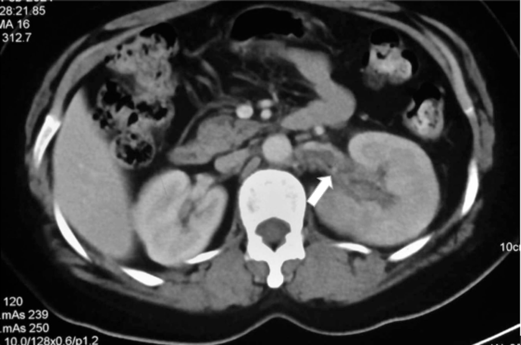
Axial CT scan image in venous phase (70 s after start of intravenous contrast injection) shows hypoattenuating eccentric filling defect in the left renal vein (thick arrow). The left kidney is enlarged, globular in appearance with reduced enhancement as compared with the right kidney. Note normally opacified right renal vein and inferior vena cava.
RVT is a rare but serious complication of many systemic diseases and should always be considered in the differentials of flank pain and haematuria.1 In adults, causes of RVT include underlying hypercoagulable state including activated protein C and protein S deficiency, antithrombin III deficiency, factor V Leiden and antiphospholipid antibody syndrome. Other causes include trauma, renal tumours, postpartum state and nephrotic syndrome.2 3 Rarely hypothyroidism is associated with procoagulant state.4 Our patient had no other predisposing factor for RVT after extensive evaluation. In the absence of specific laboratory tests, early imaging with CT angiography remains the cornerstone of diagnosis. Anticoagulation should be initiated early to prevent thrombus propagation and serious thromboembolism. Initial therapy is with heparin that is later switched to warfarin after a overlap period of 5–7 days. Duration varies from minimum of 1 year to lifelong depending on the continued presence of risk factors or recurrence. Recently, direct-acting oral anticoagulants have been used for the treatment of venous thromboembolism with good outcomes. Thrombectomy/thrombolysis should be considered in cases of bilateral RVT, thrombus extension into inferior vena cava and treatment failure while on anticoagulation.2 3
Learning points
The clinical presentation of acute renal vein thrombosis can often be confused with nephrolithiasis.
A high clinical suspicion and early imaging with CT is important to prevent extension of thrombus and prevent significant loss of renal function.
CT angiography is almost 100% sensitive and specific to diagnose renal vein thrombosis.
Ethics statements
Footnotes
Contributors MG, JS: case identification, writing and proof reading. MS: radiological image acquisition.
Funding The authors have not declared a specific grant for this research from any funding agency in the public, commercial or not-for-profit sectors.
Competing interests None declared.
Provenance and peer review Not commissioned; externally peer reviewed.