Article Text

Statistics from Altmetric.com
Description
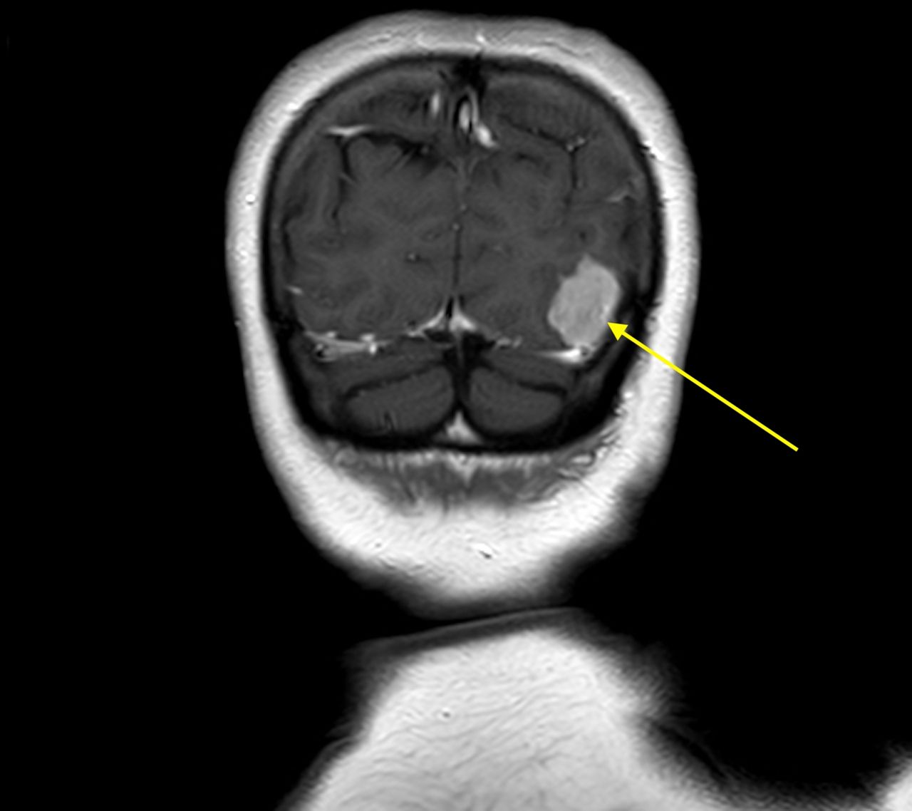
A 63-year-old woman presented with symptomatic hypercalcaemia, with a calcium level of 2.83 mmol/L (normal range 2.2–2.6 mmol/L) and an inappropriately elevated parathyroid hormone level of 14.6 pmol/L (normal range 1.6–6.9 pmol/L). Calculated urinary calcium:creatinine clearance ratio was 0.0179, in keeping with a diagnosis of primary hyperparathyroidism (PHPT). Surgical management was considered in the presence of symptoms and osteoporosis denoting end organ damage. Parathyroid scintigraphy was performed with technetium-99m sestamibi (MIBI) scan with early and late planar and single-photon emission CT (SPECT) imaging followed by ultrasound. Besides a left lower pole parathyroid adenoma, left posterior occipital lobe focal MIBI uptake was also demonstrated on the parathyroid scintigram (figure 1). CT and MRI of the brain demonstrated an extra axial, avidly enhancing 17 mm left occipital tumour diagnostic of a meningioma (figure 2). Following neurosurgical review, in the absence of neurological deficits and lack of clinical and radiological malignant features, a watch-and-wait approach was adopted.

Series of coronal SPECT delayed washout MIBI images, A anterior to C posterior. These demonstrate focal uptake within the left lower pole parathyroid adenoma (blue arrow, image A). The left occipital meningioma is seen as a focal uptake of MIBI on images B (red arrow). and C. The uptake in the salivary glands, right axilla, shoulders and neck muscles is expected physiological MIBI uptake and is not pathological. MIBI, Tc 99m sestamibi; SPECT, single-photon emission CT.

Coronal T1 cranial acquisition MRI after intravenous gadolinium demonstrating an extra-axial lesion in the left occipital region with intense homogenous enhancement confirming the occipital meningioma (yellow arrow).
Localisation of parathyroid adenomas reduces operating time and perioperative morbidity following parathyroidectomy.1 2 Parathyroid MIBI scan is the commonly used localisation technique with a reported high sensitivity and specificity of 72% and 99%, respectively.3 MIBI uptake is related to tissue perfusion, cell membrane integrity and mitochondrial activity.4 However, the increased radiotracer uptake is not exclusive to the mitochondria-rich oxyphilic cells in parathyroid adenomas. Incidentalomas can be detected with extraparathyroid and extracardiac findings in up to 27% of studies, the most frequently involving thyroid and pulmonary tumours.5 To the best of our knowledge, this is the first reported case of an incidental meningioma detected on parathyroid MIBI scintigram during localisation of parathyroid tumours.
MIBI uptake in meningiomas is the result of increased mitochondrial activity and perfusion in these tumours. The degree of radiotracers uptake including Thallium-201 and MIBI in meningioma may predict biological aggressiveness of these tumours.4 SPECT with MIBI could be used to provide prognostic information as complementary imaging tool for evaluation of meningiomas.4 Meningiomas have been reported in conjunction with PHPT both in sporadic cases6 and in association with multiple endocrine neoplasia types I and IV with mutations in the genes MEN1 and CDNK1B, respectively.7 Although not requiring current neurosurgical management, the incidental detection of meningioma in this case is clinically significant, prompting further endocrinological and genetic investigations. This report aims to raise awareness of non-parathyroid and non-cardiac incidental findings that can be easily missed. This information is important to all clinicians reviewing MIBI scintigrams.
Learning points
Clinicians need to be vigilant when reviewing technetium-99m sestamibi scans of the head and neck as incidental unrelated tumours can be visible.
Uptake of technetium-99m sestamibi is not specific for parathyroid tumours/hyperplasia and may be observed in other tumours with increased mitochondrial activity or tissue perfusion like meningioma.
Ethics statements
Patient consent for publication
Footnotes
Contributors KK and FB wrote the manuscript. KK performed the literature review. FB, CE and NA provided guidance and edited the manuscript.
Funding The authors have not declared a specific grant for this research from any funding agency in the public, commercial or not-for-profit sectors.
Competing interests None declared.
Provenance and peer review Not commissioned; externally peer reviewed.