Article Text

Statistics from Altmetric.com
Description
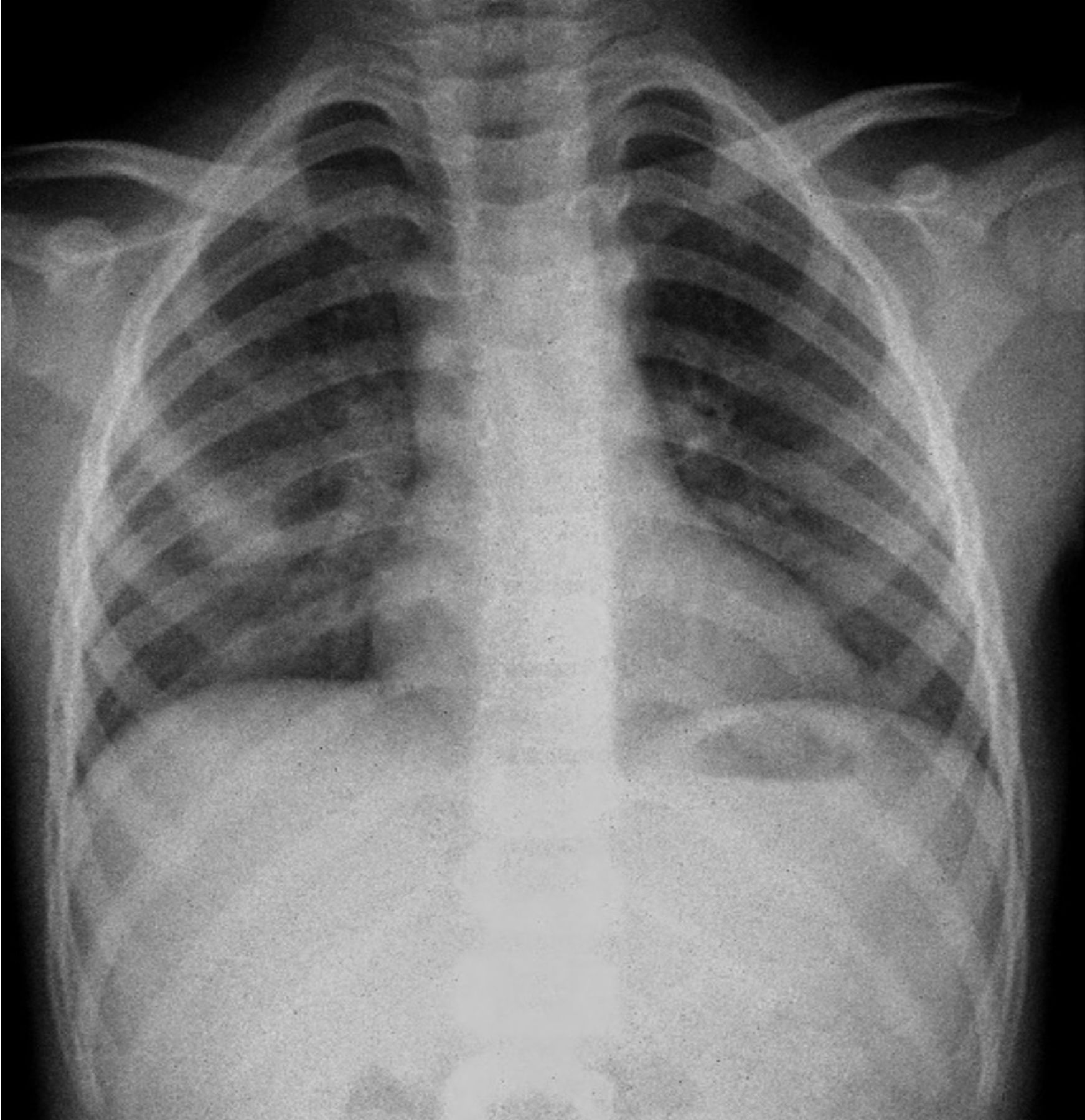
We describe the case of a previously healthy 9-year-old girl hospitalised due to hypoxaemic pneumonia. Two months after discharge, she maintained low oxygen saturations and persistent dry cough without dyspnoea, haemoptysis, migraine or palpitations. Cardiac and pulmonary auscultation was normal, and cyanosis or clubbing were not present. There was no personal or family history of mucocutaneous bleeding or telangiectasias. Thoracic radiography showed peribronchovascular thickening (figure 1). Due to persistence of symptoms, computed chest tomography (CCT) was performed and revealed pulmonary arteriovenous malformation (PAVM). Conventional angiography confirmed a large PAVM (23×19 mm) on the right parahilar topography with afferent artery draining into the right upper pulmonary vein and efferent vessel draining into the left atrium (figure 2). Percutaneous closure was performed with implantation of an Amplatzer-Duct-Occluder-I device (5/4). Clinical follow-up showed regression of hypoxaemia and she remained asymptomatic without recurrence.

Thoracic radiography showing peribronchovascular thickening.

Pulmonary angiography revealing a large pulmonary arteriovenous malformation (23×19 mm) on the right parahilar topography with afferent artery draining into the right upper pulmonary vein and efferent vessel draining into the left atrium.
PAVMs are structurally abnormal vascular communications that provide continuous right-to-left shunt between pulmonary arteries and veins. Limited prevalence data suggest that PAVMs may affect 1 out of 2600 individuals, being 1.5–2 times more frequent among women.1 Most PAVMs are congenital and due to hereditary haemorrhagic telangiectasia (HHT) but can occur in a variety of acquired medical conditions: hepatic cirrhosis, penetrating chest trauma, mitral stenosis, Fanconi syndrome, schistosomiasis, actinomycosis and metastatic thyroid carcinoma.2–8 PAVMs can also develop as a complication after congenital heart surgery.9 10 PAVMs are classified as simple or complex. Simple PAVMs are the most frequent (80%–95%) and are characteristically perfused by arteries arising from a single subsegmental artery.11 12 Complex PAVMs are perfused by more than one subsegmental artery and are typically telangiectases.13 In about 95% of cases, PAVMs are supplied by pulmonary arteries draining in pulmonary veins. Rarely they are fed by systemic arteries and/or drain into the left atrium or inferior vena cava, as we observed in our case. About 60% of cases are asymptomatic, incidentally found on chest imaging or during HHT screening.14–19 Most common symptoms are dyspnoea (13%–56%) and haemoptysis (7%–30%).14 15 18
PAVMs are typically diagnosed by CCT or conventional pulmonary angiography. Usually, complications are due to paradoxical emboli that may cause ischaemic strokes, myocardial infarction, cerebral and peripheral abscesses, discitis and migraines.17 19–23 Serious complications from PAVMs like massive haemoptysis and haemothorax are particularly more common in PAVMs>3 mm.24–26 Risk–benefit analysis in adult patients are almost always in favour of treatment.27–29 However, well-defined recommendations are still lacking for paediatric patients.
PAVM occlusion by embolisation is the gold standard of care.17 21 25 The selection of who to treat depends on factors including feeding artery diameter (FAD), PAVM-related symptoms and patient’s ability to tolerate the procedure.14 16 17 30–34 PAVMs that progressively enlarge (FAD≥2–3 mm) or become symptomatic should undergo evaluation for embolotherapy with pulmonary angiography. Surgery is an option for patients who fail repeated embolisation, for lesions suitable for intervention but not amenable for embolotherapy and for those who present with life-threatening acute haemorrhage from ruptured PAVM in a facility without access to embolotherapy.8 35–38 Due to paucity of data in children, more investigation is needed to state clear recommendations and uniformise therapeutic approach in this specific group.
Learning points
Pulmonary arteriovenous malformations (PAVMs) are rare but an important consideration in the differential diagnosis of common pulmonary signs or symptoms, such as dyspnoea or hypoxaemia.
PAVMs are typically diagnosed by computed chest tomography or conventional pulmonary angiography and their importance stems from the risks they pose (paradoxical embolic stroke, abscess, myocardial infarction and life-threatening haemorrhage).
Not all PAVMs require intervention. When indicated, most patients are treated with embolotherapy.
Ethics statements
Patient consent for publication
References
Footnotes
Contributors All authors have been personally and actively involved in substantial work leading to the paper and will take public responsibility for its content. MFC—bibliographical search, study design, data collection, analysis and interpretation of data, and drafting of the article. OM—bibliographical search, study design, data collection and critical reviewing of the content of the article. RR—bibliographical search, study design, data collection and critical reviewing of the content of the article. ARA—bibliographical search, study design, data collection and critical reviewing of the content of the article.
Funding The authors have not declared a specific grant for this research from any funding agency in the public, commercial or not-for-profit sectors.
Competing interests None declared.
Provenance and peer review Not commissioned; externally peer reviewed.