Article Text

Abstract
Monocular elevation deficiency poses a challenge to strabismus surgeons on account of its varied clinical presentations as well as management which often needs a tailored approach. We report on a young child who presented to us at 6 months of age with a clinical course marked by primary involvement of the inferior rectus muscle in one eye causing restricted elevation in all gazes and complete relief of hypotropia following disinsertion of the affected muscle but followed by recurrence and additional procedures (antimitotic application and superior rectus plication) for the same. She followed a recalcitrant clinical course which was marked by multiple recurrences requiring a tailored approach and finally managed successfully with a follow-up of 3 years, by now. This case demonstrates the almost intractable nature of restrictive pathology involving a single muscle warranting multiple surgeries and a close follow-up with good surgical outcome.
- Anterior chamber
- Visual pathway
This is an open access article distributed in accordance with the Creative Commons Attribution Non Commercial (CC BY-NC 4.0) license, which permits others to distribute, remix, adapt, build upon this work non-commercially, and license their derivative works on different terms, provided the original work is properly cited and the use is non-commercial. See: http://creativecommons.org/licenses/by-nc/4.0/.
Statistics from Altmetric.com
Background
Monocular elevation deficiency (MED) is defined as a congenital or acquired deficiency in elevation in abduction as well as adduction with varied clinical presentations like hypotropia, ptosis or pseudoptosis, reduced or normal saccadic velocity in upgaze and varied forced duction test (FDT) results depending on the underlying aetiology. MED can arise from an isolated superior rectus paresis, inferior rectus (IR) restrictive pathology or supranuclear lesions arising from pineal tumours, acoustic neuromas or acquired cardiovascular disorders like hypertension, vasculitis and thromboembolism.1 2 The onset of a large angle unilateral strabismus in early infancy and childhood poses a risk to development of normal binocular vision and needs to be addressed on priority. The management of these recurrent, restrictive cases of strabismus needs to be discussed with caregivers and a thorough evaluation is needed to rule out coexistent or causative pathologies like progressive external ophthalmoplegia, congenital fibrosis syndromes, traumatic muscle entrapment and supranuclear causes.
Case presentation
A 6-month-old female child with normal birth and developmental history presented with progressive downward deviation of right eye noticed incidentally by the parents following a fall from the bed at the age of 3 months. Family history of ptosis or eye movement anomalies was negative. There was no evidence of head or spinal injury on clinical evaluation and imaging and no evidence of orbital floor fracture on non-contrast computed tomogram (NCCT) orbit. On the clinical evaluation, there was a large right hypotropia (40 PD) without a compensatory head posture (figure 1). There was restricted elevation in abduction, primary position and adduction (−4) with normal anterior segment and fundus examination. Bell’s phenomenon was poor in the affected eye. There was no ptosis or any other synkinetic phenomena on evaluation. No horizontal deviations were noted. The child fixated with her left eye and maintained a central and steady fixation.

Preoperative photos showing large hypotropia in right eye.
Investigations
Cycloplegic refraction was done using atropine 1% eye ointment and revealed mild hyperopia. Non-contrast CT orbit showed no features of orbital fracture or muscle entrapment.
Differential diagnosis
Congenital fibrosis of the extraocular muscles is characterised by bilateral, congenital and classically non-progressive involvement of extraocular muscles causing a restrictive ophthalmoplegia which is not the case in our patient. Orbital floor fracture with IR entrapment was ruled out by NCCT orbit and clinical evaluation. Chronic progressive external ophthalmoplegia is an inherited mitochondrial cytopathy characterised by bilateral, severe, symmetric ophthalmoplegia with systemic anomalies (cardiac conduction defects) and pigmentary retinopathy with onset usually in third to fourth decade. Our patient was a child and did not have any of these clinical or imaging findings, ruling out the differential diagnoses.
Treatment
This child underwent examination under anaesthesia at this centre soon after presentation, where an FDT revealed a very tight IR muscle. An inferior limbal incision was made to facilitate adequate surgical exposure, but it was exceedingly difficult to hook the muscle. Subsequently while applying minimal traction on the hook, the IR released suddenly, with the forced duction becoming completely free for the IR, which could not be retrieved, and conjunctiva was closed. Postoperatively, resolution of hypotropia was noted in the primary position, with good elevation on first postoperative day (figure 2). At 6 weeks follow-up, there was recurrence of hypotropia (30 PD) and limited elevation (−3) (figure 3) . Exploration was done followed by identification of the IR 1 mm from the insertion site, which was then recessed by 4.5 mm along with application of antimitotic agent (mitomycin C 0.02%) for 1 min around the insertion. Postoperatively, the child maintained good alignment for 6 months, followed by recurrence of hypotropia (25 PD), increasing to 40 prism diopter in upgaze. Saccades from downgaze to upgaze were abruptly halted, suggesting restrictive pathology. Finally, a repeat IR disinsertion along with MMC application 0.02% and superior rectus plication in the right eye of 5.5 mm was done. Intraoperatively, the FDT for IR was very tight and dissection of the muscle and intermuscular septum revealed extensive fibrosis around the insertion. IR muscle was disinserted, allowing the free rotation of the globe in supraduction confirmed by FDT. Antimitotic application of MMC 0.02% was aimed at reducing the extensive fibrosis and contracture and superior rectus plication was done to minimise the chances of anterior segment ischaemia and improve the elevation. The postoperative period was uneventful, and the child had relief from hypotropia on follow-up visits (figure 4).

Postoperative photographs on day 1 showing resolution of hypotropia and improved elevation.

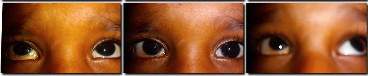
Postoperative recurrence with restricted elevation in right eye.
Postoperative photos showing complete resolution of hypotropia and good ductions in upgaze.
Outcome and follow-up
The child has been on regular follow-up in our department with good outcome. She underwent cycloplegic refraction at 2 years of age which revealed hyperopic refractive error of +2.5 DS OD and +1.5 DS OS. BCVA was 6/18 OD and 6/9 OS. Part-time occlusion therapy was started in left eye (6 hours/day) with follow-up and strict compliance to spectacle wear. She has good alignment in primary gaze as well as good ductions in up and downgaze on her follow-up visits.
Discussion
The main aim of surgery in MED is correction of hypotropia and improved elevation without unduly compromising the downgaze. The surgical management of MED is dictated by the FDT for IR muscle and recession of the same in variable amounts if restriction is noted. The main concerns with a large IR recession are the weakening of infraduction and changes in the palpebral aperture. Vertical transposition of the horizontal rectus muscles (Knapp’s procedure) and its modifications (modified or augmented Knapp’s) are indicated for correction of the large vertical deviation and improve elevation.
We reviewed existing literature on the subject to identify the unique features in our patient. Awadein and El-Fayoumi describe a surgical approach of ipsilateral moderate IR recession (up to 4 mm) with a contralateral large superior rectus recession (5–12 mm) to reduce the under action of IR and improve alignment in primary, upgaze and downgaze.3 Scott and Jackson emphasised the role of IR restriction in the pathogenesis of MED which agrees with the finding of recurrent fibrosis of IR muscle in our case.4 Metz believed that most of the cases of MED had IR restrictive pathology with superior rectus paresis being the aetiology in about 25% cases.5 We noted restriction of the IR on forced duction, with a large hypotropia in primary position without superior rectus paresis, which mirrors the observation by Metz. Wright stated the incidence of IR restriction as cause of MED in 70% cases.6 Paolillo et al reported a similar case with onset of hypotropia at 2 months of age.7 Souza-Dias et al described three cases of acquired progressive restrictive strabismus in infancy.8 One case of IR fibrosis reported by Prieto-Diaz and Laguens showed mitochondrial alterations, with possible subclinical myositis as the cause.9 The possibility of external ophthalmoplegia or congenital fibrosis of extraocular muscles in rare in our patient due to the unilaterality, lack of involvement of other muscles or progression to involve these over the follow-up period. The case reported by us is intriguing by virtue of being progressive and recalcitrant in clinical course with extremely tight IR muscle on FDT. The standout features in our patient are the early onset of hypotropia, absence of ptosis or involvement of other extraocular muscles, spontaneous improvement of hypotropia following hooking of the muscle with release of forced duction without any muscle surgery followed by recurrence which was managed by recession of the muscle with antimitotic application and finally, disinsertion of the IR with plication of superior rectus. In our case, we preferred to perform an ipsilateral superior rectus plication instead of a contralateral SR recession, with good correction of hypotropia and improved elevation without any changes in palpebral aperture height or induced torsional changes. We used mitomycin C 0.02% locally as an adjunct for its antimitotic properties in our case in view of the severe fibrosis around the muscle with good results and no ocular complications. This has been reported in literature previously by Mahindrakar et al and Chen et al with good results which mirror our result.10 11
Learning points
Always identify the underlying pattern of monocular elevation deficiency, that is. restrictive, paretic or secondary to CNS lesions preoperatively with neuroimaging or orbital imaging where warranted.
Forced duction testing for inferior rectus must be performed in all cases and recession of the muscle is warranted if positive, adjusted accordingly.
Limbal incision provides greater exposure in restrictive cases, with care while handling the tight muscles to avoid splitting or rupture.
Ciliary vessel sparing procedures like reinforced plication are recommended, especially in repeat or multiple surgeries since vertical muscle surgery has a higher chance of anterior segment ischaemia.
Ethics statements
Footnotes
Twitter @Stereoacuity@drunni81
Contributors DU and AS worked up the case and prepared the manuscript. PS operated on the case and guided the manuscript preparation.
Funding The authors have not declared a specific grant for this research from any funding agency in the public, commercial or not-for-profit sectors.
Competing interests Authors have no competing interests
Provenance and peer review Not commissioned; externally peer reviewed.