Article Text

Statistics from Altmetric.com
Description
Neonatal spontaneous arterial thromboembolism is uncommon and therefore data on risk factors, diagnostic strategies, therapeutic interventions and follow-up are scarce.1 2 Increased risk of thromboembolic events occurs in the neonatal period both due to maternal risk factors and relative immaturity of the neonatal haemostatic system.1 ,3 Most common maternal risk factors are advancedmaternal age, obesity, infections, pre-eclampsia, hypertension, caesareansection, decreased fetal movements, oligohydramnios, induction with prolongedlabour, lupus or diabetes.1 Treatment and follow-up in a tertiary reference centre is recommended, because it seems to be associated with high mortality and morbidity.1
A male neonate was born at 38 weeks gestational age from a previously healthy 33-year-old mother with a body mass index of 24 kg/m2. Pregnancy was complicated by third-trimester gestational diabetes, which required treatment with metformin.
Eutocic delivery occurred at a level II perinatal hospital, after 12 hours of labour. No sign of maternal infection was detected.
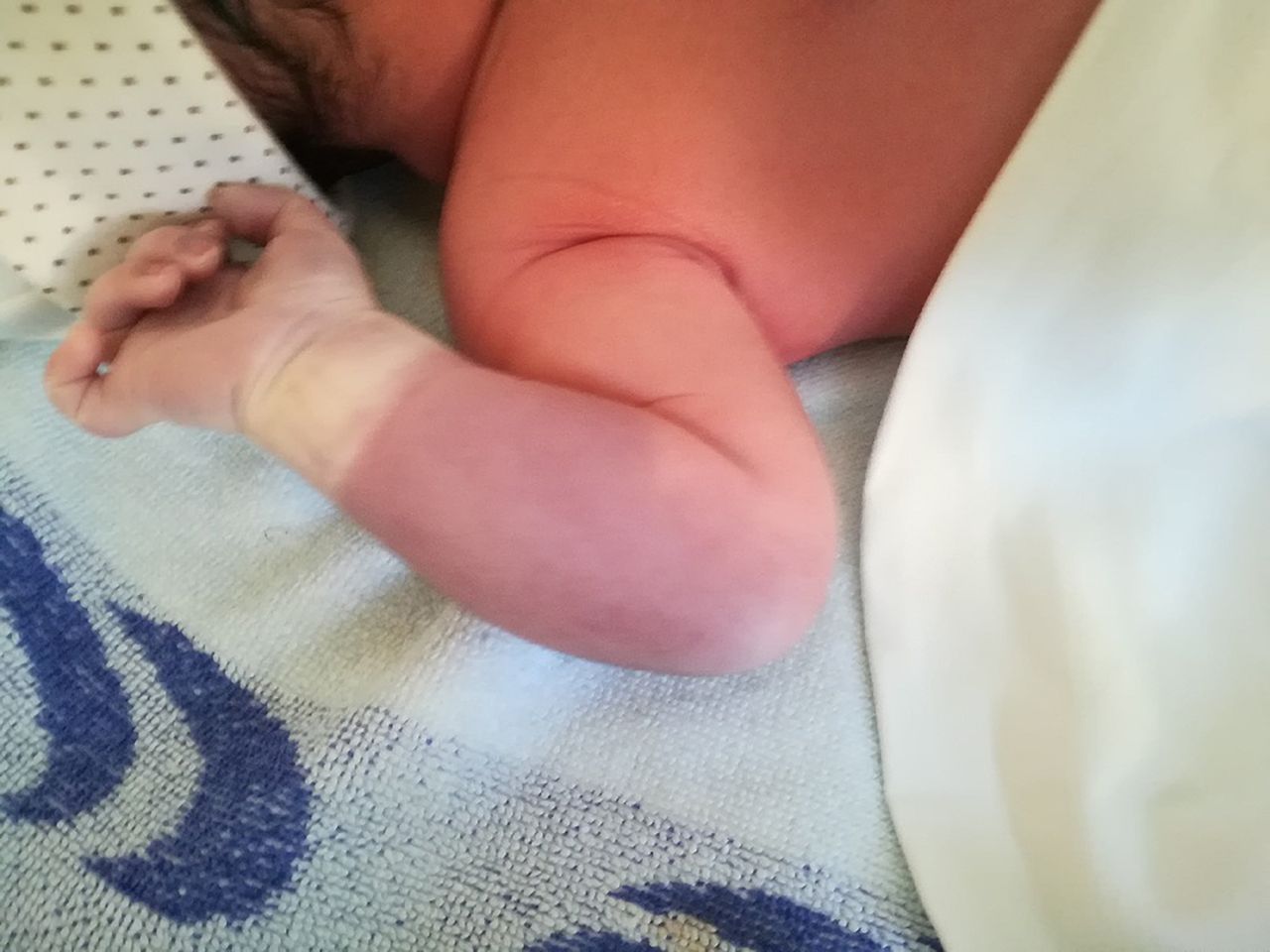
The newborn weighed 3195 g and had an APGAR index of 9 at the first minute and 10 at the fifth minute. He presented at birth with right upper limb cyanosis, pale hand (figure 1), non-palpable pulse and unmeasurable peripheral oxygen saturation. An arterial thrombotic event was suspected, and the neonate was transferred to a level III paediatric centre, where anticoagulation was initially started with unfractionated heparin, followed by low molecular weight heparin (LMWH) adjusted to anti-factor Xa levels.

Right upper limb cyanosis, pale hand, non-palpable pulse and unmeasurable peripheral oxygen saturation.
Blood tests at 24 hours of life were normal, with haemoglobin 196 g/L, haematocrit 54.5% and platelets 213×109/L . Doppler sonography revealed normal flow in the right arm arteries down to the elbow, an echogenic thrombus with 22 mm×3 mm in the deep humeral artery and absent arterial flow downstream (figure 2). At third postnatal day, the patient developed fever and early-onset neonatal sepsis was suspected with C protein reaction of 50 mg/L (reference <5 mg/L), thrombocytopaenia 50×109/L (reference 200–250×109/L). LMWH was maintained and therapy with ampicillin, gentamicin and flucloxacillin was started, with no adverse events. Blood culture was negative. There was a progressive normalisation of clinical and imaging features in the upper limb.

An echogenic thrombus with 22 mm×3 mm in the deep humeral artery.
After discharge, the infant was kept on LMWH for 4 months with complete resolution on follow-up evaluation and harmonious growth of upper limbs.
Screening for hereditary thrombophilias and inherited thrombophilia (antithrombin 3, protein C and S deficiencies, or factor V Leiden or prothrombin G20210A mutations) was excluded. Screening for cardiac or brain anomalies (low cardiac output, cord abnormalities, patent ductus arteriosus) was negative.
Other maternal risk factors, like hypertension and lupus/antiphospholipid syndrome, were excluded.
Learning points
Thromboembolic events associated with vascular catheterisation are frequently reported, but few spontaneous neonatal thromboembolism cases have been published.
Anticoagulation is the usual therapeutic approach for occlusive thrombi in the neonate. Low molecular weight heparin was preferred to unfractionated heparin due to lower haemorrhagic risk and dose adjusted according to anti-factor Xa levels.
Doppler sonography was useful for the diagnosis and therapeutic follow-up.
Ethics statements
Patient consent for publication
Footnotes
Contributors JF and AIC were responsible for the patient’s daily observation. STF was the attending physician responsible for the patient’s clinical and therapeutic orientation, namely, during sepsis. PK was the haematologist responsible for the diagnosis and follow-up after the patient’s discharge.
Funding The authors have not declared a specific grant for this research from any funding agency in the public, commercial or not-for-profit sectors.
Competing interests None declared.
Provenance and peer review Not commissioned; externally peer reviewed.