Article Text

Statistics from Altmetric.com
Description
A 35-year-old female patient reported with a problem of bleeding and swollen gums in the lower front tooth region for 2 weeks associated with mild pain, which is intermittent and aggravates on consuming any type of food.
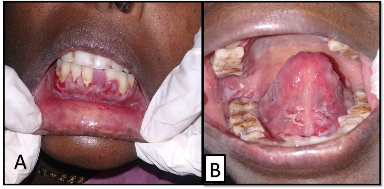
Intraoral examination revealed multiple ulcerations and erosions with crustations seen on right and left buccal mucosa, floor of the mouth and gingiva. Ulcers presented with the well-defined borders with yellowish slough with the areas of reddish erythema. Ulcers were tender on palpation and not attached to the underlying structures (figure 1A,B). Based on the clinical presentation, provisional diagnosis of pemphigus vulgaris was given. Incisional biopsy of gingiva was carried out and report revealed hyperplastic striae with squamous parakeratinised epithelium with underlying connective tissue stroma. Hyperbasilar clefting was seen with large eosinophilic acantholytic cells (Tzanck cells) with basophilic nucleus. Basement membrane was intact with connective tissue stroma, which was highly cellular in nature with proliferating blood vessels and fibrinous exudate (figure 2). Considering all the clinical and histopathological findings, a final diagnosis of pemphigus vulgaris was given.

(A) Erosions seen on the gingiva, which is soft and edematous. (B) Erosions seen on the floor of the mouth and ventral aspect of tongue.

Histopathological sections revealing Tzanck cells.
Pemphigus vulgaris is the most common form of pemphigus, accounting for over 80% of cases. In most patients, it affects the oral mucosa and is sometimes difficult to diagnose when only mucosal involvement is present.1 Desquamative gingivitis is the most common manifestation of the disease when gingival is involved, as reported in our case.2
Treatment is usually targeted at controlling the severity and preventing relapses. Systemic corticosteroids remain the gold standard treatment for pemphigus. It is preferable to start with low doses of prednisolone (0.5–1.5 mg/kg/day) initially. If there is no adequate response, then dose may be increased to up to 2.0–2.5 mg/kg/day.3
Dental and periodontal follow-up is very necessary after attaining clinical remission, at least two to three times a year for the first year following diagnosis.4
Learning points
Pemphigus vulgaris is the most common variant that affects the oral cavity.
Oral lesions are the first manifestation of the disease in 50%–90% of the cases.
The treatment should be organised by the dermatologist along with close cooperation of periodontologist for oral treatment.
Footnotes
Contributors VN contributed to conception and design, acquisition of data or analysis, interpretation of the data, drafting the article or revising it critically for important intellectual content, final approval of the version published and agreement to be accountable for the article and to ensure that all questions regarding the accuracy or integrity of the article are investigated and resolved. RK and PKR contributed to revising important intellectual content. US contributed in the final approval.
Funding The authors have not declared a specific grant for this research from any funding agency in the public, commercial or not-for-profit sectors.
Competing interests None declared.
Patient consent for publication Obtained.
Provenance and peer review Not commissioned; externally peer reviewed.