Article Text

Statistics from Altmetric.com
Description
A 39-year-old man with necrotising pancreatitis was admitted to the intensive care unit (ICU). On day 14, his right-sided central venous catheter (CVC) was replaced with a left-sided catheter. A confirmatory chest X-ray revealed that rather than charting its usual course across the left brachiocephalic vein into the superior vena cava (SVC), the catheter journeyed straight down into the left atrium (figure 1). A CT thorax (figure 2) revealed the patient had two SVCs, and the patient’s left-sided SVC had been unintentionally catheterised.
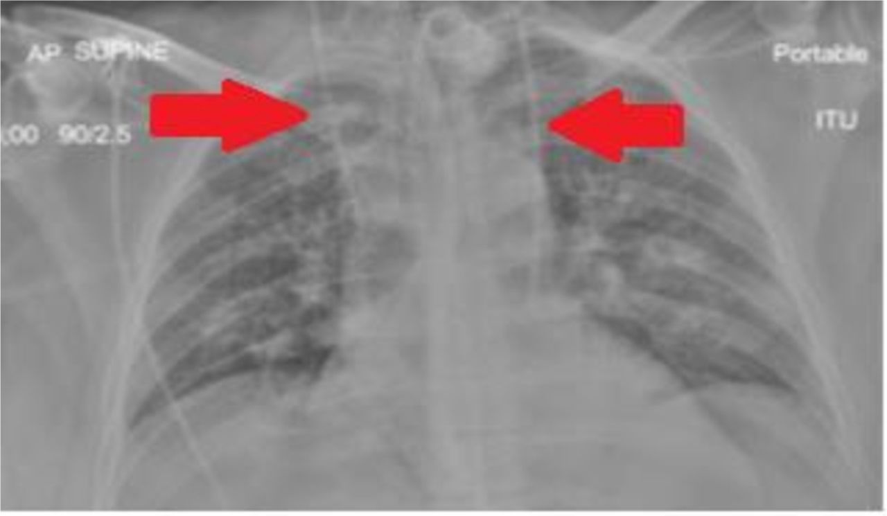
Anteroposterior chest X-ray (AP CXR) shows the left-sided CVC tracking into the left atrium. The right SVC is already catheterised. CVCs are indicated by the red arrows. CVC, central venous catheter; SVC, superior vena cava.

CT thorax shows two superior vena cavae as indicated by red arrows.
A percentage range of 0.3%–0.5% of the population have two SVCs or a persistent left SVC (PLSVC). There is an increased (1.3%–4.5%) prevalence of PLSVC in patients with congenital heart disease.1 It occurs due to failure of the left anterior cardinal vein to obliterate during fetal development and is an incidental finding in the vast majority of cases. Ninety-two per cent of PLSVCs drain into the coronary sinus, but the remaining 8% drain into the left atrium causing a right-left shunt which is usually insignificant.2 However, as a result of the latter group, central catheters pose a risk of systemic embolisation so should be removed if the patient’s cardiothoracic anatomy has not been formally assessed. If there is no shunt present, catheters can remain though with due care and attention to the nearby coronary sinus and the associated risk of arrhythmias from a misplaced catheter tip.
In this case, the exact position of drainage from the extra SVC was uncertain so the CVC was replaced with another right-sided catheter. The patient was eventually discharged from the ICU and went on to make a full recovery.
Learning points
Consider a persistent left superior vena cava (PLSVC) when a left-sided central venous catheter appears in an unusual position in the left thorax.
Left-sided superior vena cava catheters can remain in situ though only if formal imaging (eg, echocardiography) of the cardiothoracic anatomy confirms the absence of a shunt.
PLSVC is often associated with congenital cardiac abnormalities.
Footnotes
Contributors JAG, a senior house officer working in intensive care, inserted the central line and found the unusual course on chest X-ray before enlisting the help of consultants to discover what had occurred. A CT thorax revealed the presence of two superior vena cavae (SVC) and the fact that the left-sided SVC had in fact been cannulated originally. KS was JAG’s clinical supervisor at the time and supervised the writing of the report—suggesting changes where appropriate. We hope this case report educates medical and nursing staff with respect to the potential dangers of left-sided SVC cannulation.
Funding The authors have not declared a specific grant for this research from any funding agency in the public, commercial or not-for-profit sectors.
Competing interests None declared.
Provenance and peer review Not commissioned; externally peer reviewed.
Patient consent for publication Obtained.