Article Text

Abstract
Puckering of the skin following a fracture is a rare sign which can result in skin necrosis if the fracture is not urgently reduced. Skin puckering is associated with humeral, tibia and clavicular fractures. We present a case of a 79-year-old woman who fell on to her outstretched hand sustaining a right radial fracture with obvious skin puckering. Following X-rays, local anaesthetic was given and the skin was reduced, the fracture manipulated and a full cast applied. The patient made a full recovery. To our knowledge, this is the first reported case of the pucker sign in an adult radial fracture.
- orthopaedics
- radiology
- orthopaedic and trauma surgery
This is an open access article distributed in accordance with the Creative Commons Attribution Non Commercial (CC BY-NC 4.0) license, which permits others to distribute, remix, adapt, build upon this work non-commercially, and license their derivative works on different terms, provided the original work is properly cited and the use is non-commercial. See: http://creativecommons.org/licenses/by-nc/4.0/
Statistics from Altmetric.com
Background
The pucker sign is a rare sign in which dimpling of the skin is associated with the development of a subcutaneous haematoma following a bone fracture in the extremities.1 The clinical sign was first reported in a proximal humeral fracture.2 Skin puckering is associated with marked displacement, soft tissue interposition and can result in soft tissue necrosis if the fracture is not immediately reduced.3 Traditionally, skin puckering has been associated with tibia, clavicle and supracondylar humeral fractures.4 The mechanism of the skin puckering in a radial fracture is similar to that in a humeral fracture: displaced radial bone penetrates surrounding soft tissue, muscle and the dermal layer of the skin, resulting in tethering of the soft tissue and interposition into the fracture site creating a near-open fracture.1 Distal radial fractures are among the most common types of fractures.5 To the best of our knowledge, this is the first case report of a pucker sign in a distal radius fracture in an adult.
Case presentation
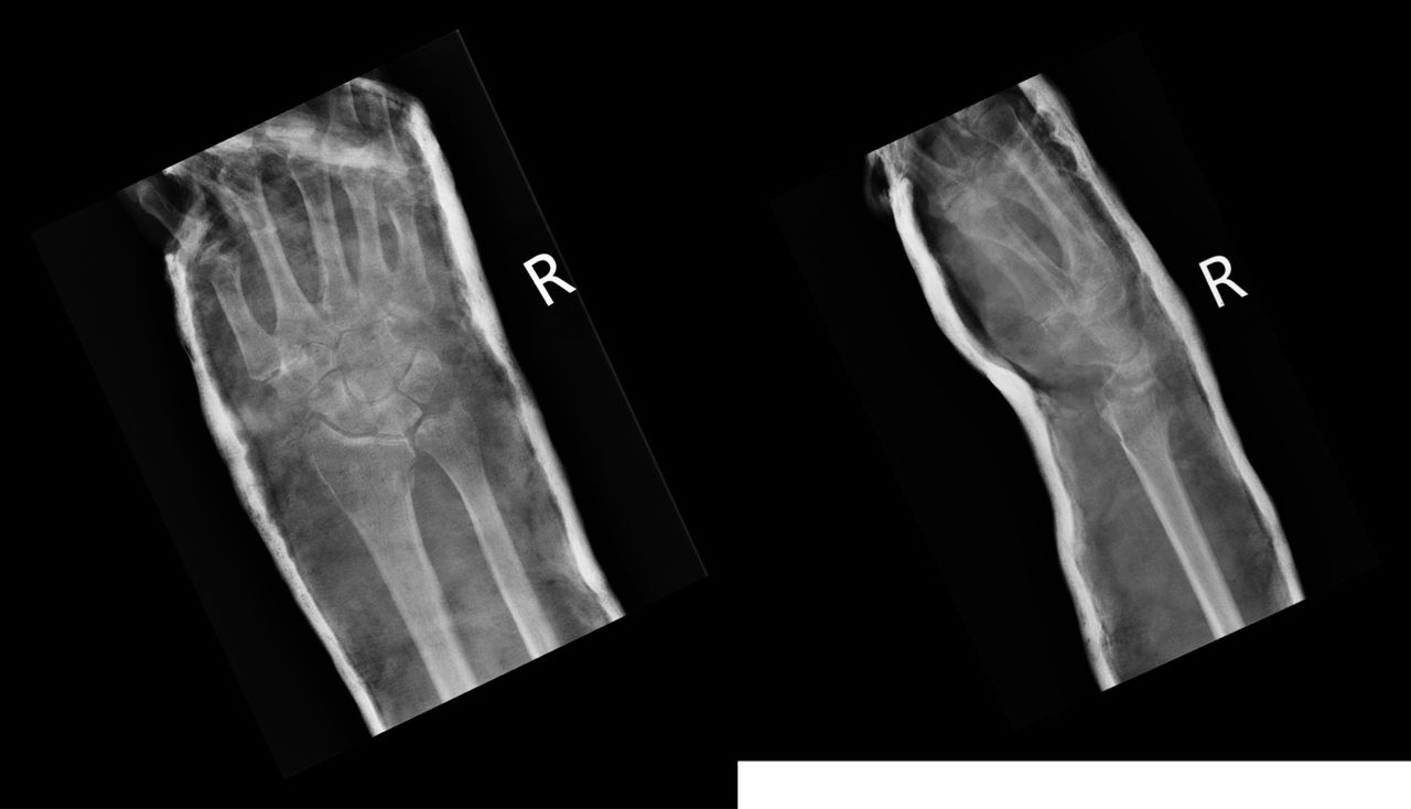
A 79-year-old woman presented to accident and emergency department following a mechanical fall on to her right outstretched hand. The patient was intact on neurovascular examination with normal power. The wrist was swollen with pucker sign over the fracture site (figures 1 and 2). X-rays taken showed a displaced fracture of the right distal radius (figure 3). Local anaesthetic was given. The skin was reduced, and the fracture manipulated with a full cast applied (figure 4). After 2 days, the cast was changed to a fibreglass cast and check X-rays were taken. Alignment was satisfactory, as such, conservative management was continued. Medical history includes atrial fibrillation, mitral and tricuspid valve repair, mild left ventricular systolic dysfunction, hypertension and a previous right neck of femur fracture in 2016 and subsequent hemiarthroplasty. The patient had no known drug allergies. Drug history includedwarfarin, digoxin, furosemide, ramipril, spironolactone, bisoprolol, atorvastatin and citalopram. The patient lived alone, was mobile with a stick and had a history of recurrent falls. She was a non-smoker and did not drink alcohol.

Pucker sign over the fracture site.

Further view of pucker sign over the fracture site.
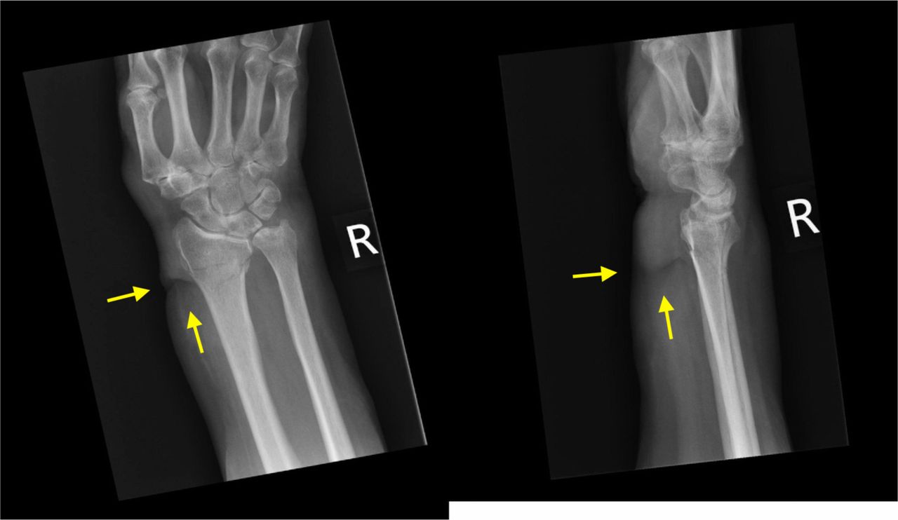
Left: anteroposterior X-ray, distal radius fracture and skin puckering. Right: lateral X-ray (at presentation to emergency department).

Left: anteroposterior X-ray postmanipulation and cast application. Right: lateral X-ray (postreduction in emergency department).
Outcome and follow-up
Two days following injury, the patient was seen in the fracture clinic. The cast was changed to a lighter fibreglass cast and anteroposterior (AP) and lateral check X-rays showed acceptable fracture position. The patient was planned to be reviewed again in 1 week’s time with AP and lateral X-ray of the right wrist on arrival. The repeated X-rays continued to show a reasonable alignment was maintained. She had a good range of movement in her fingers, the thumb was pain free, and the plaster was fitting well. After her second follow-up appointment, it was arranged for her to have a review appointment at 6 weeks from injury with her plaster off on arrival. At her 6 weeks follow-up, there was no tenderness at the fracture site. Extensor pollicis longus (EPL) was working, and she had a reasonable range of wrist and small joints mobility. She was taught some exercises to be done by herself at home to get the full movements back. The patient was reassured and discharged back to the care of her general practicioner.
Discussion
To the best of our knowledge, this is the first case of pucker sign in an adult distal radius fracture. Since the skin reduction and manipulation of the fracture, the patient has made a full recovery. The importance of this report lies in evidencing skin puckering in distal radial fractures despite being more traditionally associated with humeral fractures. Pucker sign was first reported by Alshryda et al in a humeral neck fracture.2 Since then, the literature has outlined this clinical sign and its outcome in paediatric humeral fractures.1 6–8 Only one prior case report exists of skin puckering in a distal radius fracture; however, this is of a solitary paediatric case in a skeletally immature 10-year-old boy, whereas, ours is of a fragility fracture resulting from a fall from standing height in an elderly woman.1 We present a new clinical sign in adult distal radial fractures which may aid in management. This case report implies that it provides evidence of successful management of this fracture with the presence of this clinical sign which may aid in future similar presentations as well as increasing knowledge.
Learning points
Skin puckering is associated with the development of a haematoma following a bone fracture in the extremities.
This is the first case of pucker sign in an adult distal radius fracture.
Skin puckering nearly always signifies soft tissue interposition, thus, indicating the need for manipulation and possibly, open reduction.2
Footnotes
Contributors MMA-S conceived of the presented case report. Both authors developed the structure of the case report. MMA-S encouraged CR to further investigate regarding the pucker sign and supervised the findings of this case report.
Funding The authors have not declared a specific grant for this research from any funding agency in the public, commercial or not-for-profit sectors.
Competing interests None declared.
Provenance and peer review Not commissioned; externally peer reviewed.
Patient consent for publication Obtained.