Article Text

Summary
We report a 3-month-old girl who presented with high-grade fever for 3 days. Her initial physical examination was normal. Investigation showed abnormal white cells in her urine. She was diagnosed with a urinary tract infection and received an antibiotic for 1 day. After that, she developed a generalised maculopapular rash over her body. An adverse drug reaction from the antibiotic was suspected, and the patient was referred to our hospital. On admission, she still had fever and was irritable. She was diagnosed with sepsis and given another broad-spectrum antibiotic for 2 days. However, her fever still persisted. An additional thorough physical examination showed redness of her BCG inoculation scar. Consequently, a diagnosis of Kawasaki disease (KD) was made. After she received intravenous immunoglobulin, her fever diminished straight away. This case highlights an unusual manifestation of KD in an uncommonly young age group.
- vasculitis
- paediatrics
This is an Open Access article distributed in accordance with the Creative Commons Attribution Non Commercial (CC BY-NC 4.0) license, which permits others to distribute, remix, adapt, build upon this work non-commercially, and license their derivative works on different terms, provided the original work is properly cited and the use is non-commercial. See: http://creativecommons.org/licenses/by-nc/4.0/
Statistics from Altmetric.com
Background
The usual incidence of Kawasaki disease (KD) is from 6 months up to 5 years of age.1 There are only a few reported cases of neonatal KD.2 An important and distinctive clinical sign that is not included in the classical clinical criteria of KD is a reaction at the BCG inoculation site. BCG redness is found in more than 50% of KD patients aged less than 12 months.3 The crust formation or redness at the BCG scar should alert the physician to consider KD as a differential diagnosis for any febrile children, even when there are only a few or no classic clinical presentations.4
Since it is challenging to establish a diagnosis, coronary artery complications occur frequently in infants with KD.5 To prevent such harmful consequences, paediatricians can treat patients with KD through intravenous immunoglobulin (IVIG) infusion within 10 days of onset of the disease.6 IVIG can decrease the risk of coronary artery abnormalities from 25% to 5%.5 Our patient was diagnosed rapidly enough and received IVIG in good time, so she had no residual coronary artery complications.
Persistent fever can be caused by infectious and non-infectious diseases. Infectious diseases may be caused by bacteria, viruses, fungi or parasites. In this case, we gave her broad-spectrum antibiotics, but her symptoms did not improve indicating that bacterial infection was less likely. We therefore also included other micro-organisms and non-infectious diseases such as autoimmune diseases or neoplasm in the differential diagnosis. KD should also be considered as a differential diagnosis in cases of persistent fever especially in children under 5 years. A detailed history and physical examination are required to reveal the cause of persistent fever.
Case presentation
A 3-month-old infant from northern Thailand presented with fever for 3 days. She was a healthy term infant. She received BCG vaccination at birth. Her vaccination was completed according to Thailand’s national immunisation programme. Before she was referred to our hospital, she was admitted to a primary care hospital due to high-grade fever together with poor intake. Investigations for the source of the infection were conducted. Her urine examination showed a concentration of white cells of 20–30 cells/high-power field (hpf). As a result, she was diagnosed with a urinary tract infection. She was given ceftriaxone. After that, she developed a generalised maculopapular rash, so a ceftriaxone allergy was suspected. She was referred to our hospital.
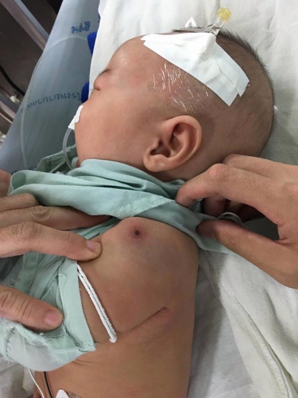
On the first day of admission to our hospital, physical examination revealed high-grade fever (39˚C), a heart rate of 155 beats per minute and a respiratory rate of 32 breaths per minute. She looked irritable. She had erythematous lips and a mildly injected pharynx. She had no erythema of the palms or soles. Other systems were within normal limits. We thought of sepsis, so we gave her meropenem. However, the fever still persisted after 2 days of the antibiotic. We repeated the physical examination and found that she had redness and induration around her BCG scar (figure 1). She had neither conjunctival injection nor cervical lymphadenopathies.

Redness and induration around her BCG scar.
Investigations
A complete blood count revealed a haemoglobin concentration of 10.7 g/dL, haematocrit of 32.1%, a mean corpuscular volume of 83.1 fL and a white cell count of 23.7 x 10⁹/L with 66% neutrophils, 22% lymphocytes and 3% monocytes. The platelet count was as high as 450 x 109/L. The erythrocyte sedimentation rate (ESR) was 19 mm/hour. C reactive protein (CRP) was elevated to 130.7 mg/L. The liver function tests revealed a total bilirubin of 0.3 mg/dL, direct bilirubin of 0.1 mg/dL, aspartate aminotransferase of 38 U/L, alanine transaminase of 21 U/L, alkaline phosphatase of 111 U/L, total protein of 5.3 g/dL, albumin of 3.3 gm/dL and globulin of 2 g/dL. Urinary analysis showed a white cell count of 20–30 cells/hpf with 1–2 epithelial cells/hpf. The cerebrospinal fluid profile was normal. All cultures from catheterised urine, blood and cerebrospinal fluid were negative.
Echocardiography showed no anatomical cardiac defect. The coronary arteries were normal sized: the left main coronary artery was 1.7 mm, left anterior descending artery 1.7 mm, left circumflex artery 1.2 mm and the right coronary artery 1.8 mm. There was normal left ventricular function and trivial mitral valve regurgitation without pericardial effusion.
Differential diagnosis
Incomplete KD (two clinical criteria of red lips and polymorphous exanthem). On additional physical examination, erythema induration at BCG inoculation site plus three supplementary laboratory criteria of white cells ≥15 000 cells/mm3, urine white cell count ≥10 cells/hpf and platelet count ≥450 000 cells/mm3).
Treatment
The patient received IVIG 2 g/kg and aspirin 80 mg/kg/day on her seventh day of fever.
Outcome and follow-up
After administration of IVIG and aspirin, the fever dramatically defervesced in the following 24 hours. The infant was discharged 48 hours later. Her vaccination programme was postponed for the next 11 months to avoid live attenuated vaccines. Aspirin was prescribed at an antithrombotic dose for 8 weeks.
A follow-up echocardiography showed normal coronary arteries. The right coronary artery was 1.2 mm, the left main coronary artery was 2 mm and the left anterior descending artery was 1.5 mm. At follow-up examination, she had no periungual peeling of fingers or toes.
Discussion
Only about 10% of KD occurs in infants who are less than 6 months.5 Moreover, the incidence of KD in infants less than 3 months in Japan and Korea is only 1.67% and 2.2%, respectively.7 8 The diagnosis of KD in infants 3 months of age or younger is difficult because very few cases (about 24%) meet 4 out of the 5 classical clinical criteria: changes in lips and oral cavity, polymorphous exanthem, bilateral non-exudative bulbar conjunctivitis, changes in extremities and a cervical lymphadenopathy over 1.5 cm in size.7 As a consequence, cardiac complications are more common in KD patients less than 6 months of age.9 IVIG and aspirin remain the mainstay of KD treatment.10 Prompt diagnosis and administration of IVIG within 10 days, or ideally before day 7 of the disease, is mandated in order to reduce such cardiac complications.11 Aspirin in the acute inflammatory period is prescribed at either 80–100 mg/kg/day or 30–50 mg/kg/day. Forty-eight to seventy-two hours after cessation of fever, aspirin should be decreased to a low dose (3 to 5 mg/kg/day). Additional therapy may include corticosteroid, infliximab and etanercept.10
Since diagnosing KD in infants younger than 6 months is difficult, any febrile infants who have fever for 7 days or more without other explanations, even without any clinical clues of KD, should receive a blood analysis of systemic vascular response. If the ESR or CRP is elevated, echocardiography should be performed.12 However, there are many limitations of echocardiography in the diagnosis of KD. First, it is an operator-dependent imaging. Second, it also requires co-operation from the patient. Third, the growth of children and the increasing body size cause difficulties in visualising the coronary arteries. These could possibly be diagnostic limitations as the sensitivity and specificity of echocardiography to determine coronary artery stenosis are uncertain.10
There are many studies concentrating on the early diagnostic criteria of infantile KD.9 13–16 Kang et al reviewed medical records of 64 KD patients from January 2010 to October 2014. Twenty of the analysed KD patients were infants less than 1 year of age. They discovered that infants had higher rates of inflammation at the BCG inoculation site (P<0.001), but lower incidence of changes in the extremities (P=0.029) and cervical lymphadenopathy (P=0.006). They stated that BCGitis is an initial sign that could lead to the diagnosis of incomplete infantile KD.13 Yoon et al reviewed medical histories of 239 KD patients from January 2013 until June 2015, of which 26 were less than 6 months. They recognised that infants less than 6 months with KD rarely express cervical lymphadenopathy (P=0.005) or non-exudative conjunctival injection (P=0.027) when compared with KD patients aged 6 months or older.9 Limbach and Lindinger claimed that all infants with KD shared similar characteristics of persistent fever despite administration of antibiotics and a polymorphous skin manifestation. They also revealed that abnormal urinalysis and thrombocytosis were frequently observed in this age group.16
In our case, the patient was only 3 months. She had BCGitis, polymorphous exanthem, persistent fever even after infusion with several antibiotics, a sterile pyuria and thrombocytosis which provided the clues for a diagnosis. She had neither cervical lymphadenopathy, changes in extremities nor conjunctival injection. Fortunately, IVIG was given within 10 days of the onset of the disease and so she had no coronary artery complications.
Learning points
Kawasaki disease (KD) should be considered in infants less than 6 months even though the incidence is low in this age group.
Careful physical examination is important. If BCGitis is found in febrile children without a source of infection, consideration of KD is warranted.
Early diagnosis of KD and administration of intravenous immunoglobulin could reduce the risk of coronary artery complications.
References
Footnotes
Contributors AW: picked a topic, made an outline and revised the manuscript. NL and AW: researched and wrote the paper.
Funding The authors have not declared a specific grant for this research from any funding agency in the public, commercial or not-for-profit sectors.
Competing interests None declared.
Patient consent Gaurdian consent obtained.
Provenance and peer review Not commissioned; externally peer reviewed.