Article Text

Statistics from Altmetric.com
Description
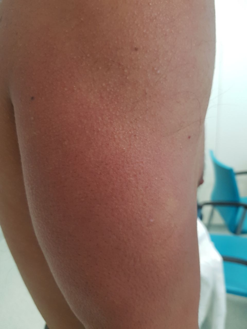
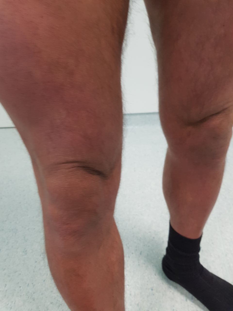
A 43-year-old previously healthy man presented for a first medical appointment at our immunology clinic with a 1 year history of painless nose and ear oedema and erythema, with no other symptoms. He had been diagnosed with relapsing polychondritis after laboratory studies revealed normal inflammatory markers, and was unsuccessfully treated with systemic corticosteroids, along with methotrexate in the last 3 months of treatment. Physical examination revealed deep furrows of the glabella (figure 1), skin thickening of the glabella, nose and ears with ear lobe involvement (figures 1 and 2), multiple 1–3 mm diameter firm papules in the limbs (figure 3) and ‘Shar-Pei sign’ in the thighs (figure 4). Lab tests showed an IgG kappa monoclonal gammopathy and normal thyroid tests. Skin biopsy revealed mucin deposition and fibroblast proliferation.

Skin thickening of the nose and glabella with deep furrows of the latter.
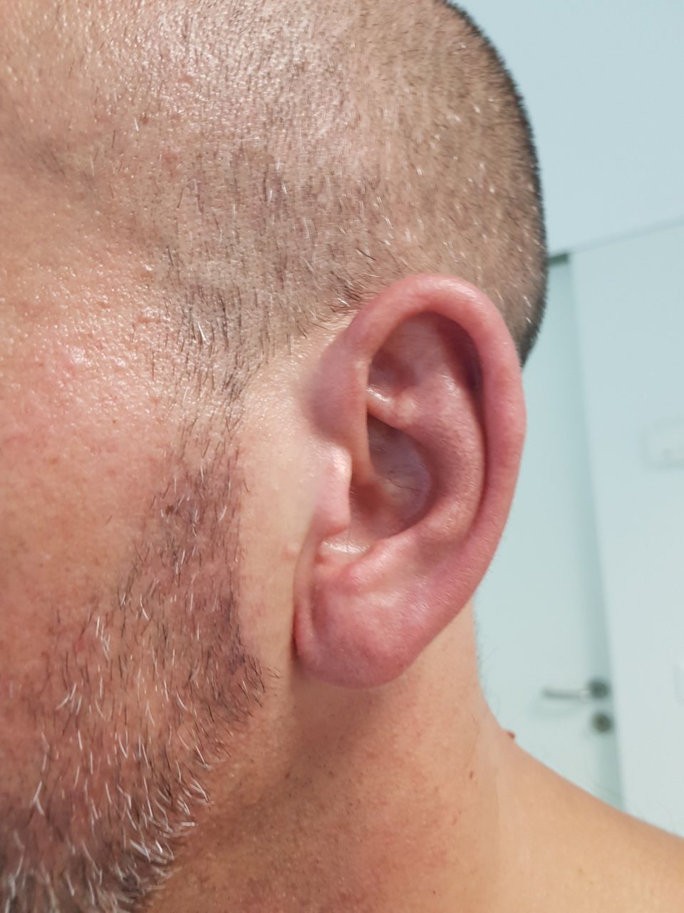
Skin thickening of the ears with involvement of the ear lobe.

Multiple 1–3 mm diameter firm papules in the limbs.

Deep furrowing of the thighs—‘Shar-Pei sign’.
The diagnosis of scleromyxoedema was made and the patient was started on intravenous immunoglobulin (two cycles of 40 g/day during 3 days) and thalidomide (50 mg/day), with significant improvement after 2 months. Therapy with immunoglobulin was kept but thalidomide was reduced due to side effects (hypersomnia and weight gain).
The differential diagnosis of scleromyxoedema includes localised lichen myxoedematosus, scleroderma and hypothyroidism-associated myxoedema as they share common dermatologic manifestations. In this case, the distribution of the lesions with glabella involvement, ‘Shar-Pei sign’ in the thighs and firm papules in the limbs, along with a monoclonal gammopathy and normal thyroid function are typical of scleromyxoedema.
Learning points
This is a rare, chronic and progressive disease of unknown origin characterised by a generalised papular eruption and sclerodermoid induration, with histopathological features of mucin deposition and fibroblast proliferation Monoclonal gammopathy is present in most cases.
Scleromyxoedema, as well as relapsing polychondritis, can involve the ear and nose. Absence of pain and involvement of the ear lobe, however, are suggestive of the first diagnosis (as seen in this case).
A correct diagnosis is crucial, since it is a chronic disease with potential systemic involvement and associated with high morbidity and mortality.
Footnotes
Contributors All authors contributed substantially to the conception of this manuscript. SP, JS and AMo were responsible for reporting the case. SP, JS and AMa wrote the learning points and performed the bibliographic research. SP had full responsibility for the final work.
Funding The authors have not declared a specific grant for this research from any funding agency in the public, commercial or not-for-profit sectors.
Competing interests None declared.
Patient consent Obtained.
Provenance and peer review Not commissioned; externally peer reviewed.