Article Text

Statistics from Altmetric.com
Description
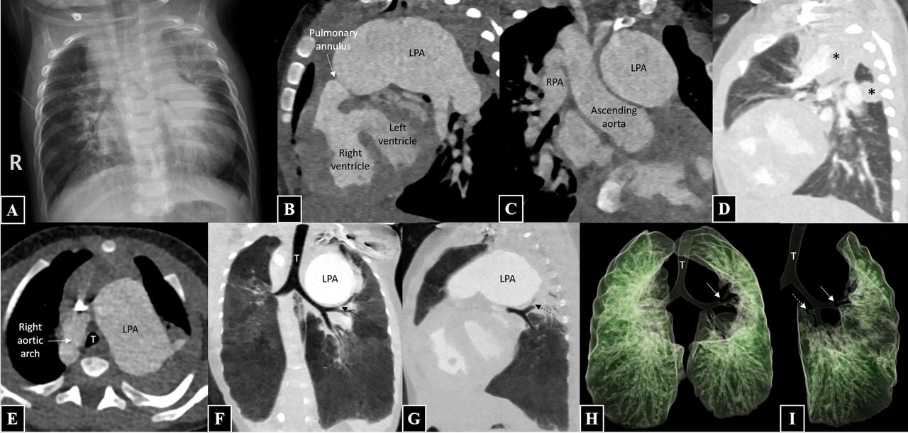
A 2-month-old boy presented to the paediatric cardiology department with cyanosis and feeding difficulties. Chest radiograph revealed cardiomegaly, right-sided aortic knuckle and dilated pulmonary artery segment along with mild indentation on the lower trachea and diffusely narrowed left main bronchus (figure 1A). A diagnosis of tetralogy of Fallot (TOF) with absent pulmonary valve syndrome was made on transthoracic echocardiogram; however, origin of right pulmonary artery (RPA) was not well visualised. The patient further underwent CT angiography (CTA) to delineate the cardiac as well as any extracardiac abnormalities.
Frontal chest radiograph (A) reveals cardiomegaly, right-sided aortic knuckle and dilated pulmonary artery segment along with mild indentation on the lower trachea and diffusely narrowed left main bronchus. Sagittal oblique maximum intensity projection (B) shows presence of features of tetralogy of Fallot, that is, right ventricular hypertrophy, perimembranous ventricular septal defect and pulmonary annular stenosis. Pulmonary valve leaflets are not seen with aneurysmal dilatation of the main and left pulmonary artery (LPA). Coronal oblique maximum intensity projection (C) shows right pulmonary artery (RPA) arising from the mid-ascending aorta. Sagittal multiplanar reconstruction (D) reveals collapse (*) of the apicoposterior segment of the left upper lobe and superior segment of the right lower lobe. Axial image at the level of arch (E) shows ‘inverted-V’-shaped trachea between the right aortic arch and the dilated LPA. Coronal multiplanar reconstruction lung window (F) reveals diffuse mild narrowing of the left main bronchus due to compression by the dilated LPA with near-complete occlusion of the apicoposterior branch of the left upper lobe bronchus (black arrow). Oblique sagittal multiplanar reconstruction lung window (G) reveals near-complete occlusion of the superior segmental branch of the left lower lobe bronchus (dotted arrow). Virtual bronchography images (H, I) show near-complete occlusion of the apicoposterior branch of the left upper lobe bronchus (black arrow) and the superior segmental branch of the left lower lobe bronchus (dotted arrow).
CTA revealed features of TOF with pulmonary annular stenosis. Pulmonary valve leaflets were absent with aneurysmal dilatation of the main and left pulmonary artery (LPA) (figure 1B). The RPA was not seen in continuation with the main pulmonary artery; instead, it was seen to arise from the mid-ascending aorta and supplying the right lung (figure 1C). Review of the lung window images revealed collapse of the apicoposterior segment of the left upper lobe and superior segment of the right lower lobe (figure 1D). Subsequent careful examination of the airway depicted an ‘inverted-V’-shaped trachea with diffuse mild narrowing of the left main bronchus, near-complete occlusion of the apicoposterior branch of the left upper lobe bronchus and the superior segmental branch of the left lower lobe bronchus, due to compression by the dilated left pulmonary artery (figure 1E–G). These findings were exquisitely demonstrated on virtual bronchographic images (figure 1H,I).
Hemitruncus is a rare defect, accounting for only 0.12% of all congenital cardiac defects. It is best described as one branch of pulmonary artery originating from the ascending aorta with the other branch coursing normally from the main pulmonary artery, which is in continuation with the right ventricle. Moreover, in TOF, anomalous origin of LPA is more common as compared with that of RPA. Similarly, absent pulmonary valve syndrome (APVS) is also seen in only about 3%–6% of all cases of TOF.1 2 Coexistence of both anomalies in the setting of TOF is understandably extremely rare. In the presence of this complex anomaly, both lungs are exposed to high volume and pressure load; the right lung will receive blood from the aorta at a high systemic pressure while the left lung receives the entire right ventricular output.3 A right heart catheterisation evaluating the pulmonary vascular resistance and its reversibility would help decide amenability to surgery. Also, APVS results in aneurysmally dilated pulmonary arteries, which often cause significant airway compression that can be accurately depicted on CT, especially reconstructed virtual bronchography images.
Early surgical repair should be performed to prevent development of irreversible pulmonary hypertension. A variety of surgical manoeuvres have been employed for repairing the artery with anomalous origin, including direct end-to-end anastomosis, interposition with a synthetic graft or a homograft patch and use of aortic flap. Concurrent complete repair of TOF with homograft placement between the right ventricle and main pulmonary artery may be performed. The aneurysmally dilated arteries may require a reduction angioplasty.
Learning points
Coexistence of tetralogy of Fallot with absent pulmonary valve syndrome and anomalous origin of right pulmonary artery from ascending aorta is extremely uncommon.
In this condition, both lungs are exposed to high volume and pressure load as the right lung receives blood from the aorta at a high systemic pressure while the left lung receives the entire right ventricular output.
Absent pulmonary valve syndrome results in aneurysmally dilated pulmonary arteries, which often cause significant airway compression that can be exquisitely depicted on virtual bronchography images.
Footnotes
NNP, AS and SK contributed equally.
Contributors NNP has participated sufficiently in the conception of the idea, development of the intellectual content, design, writing and final approval of the manuscript. AS has participated sufficiently in the conception of the idea, development of the intellectual content, design, writing and final approval of the manuscript. SK has participated sufficiently in the conception of the idea, development of the intellectual content, design, writing and final approval of the manuscript.
Funding The authors have not declared a specific grant for this research from any funding agency in the public, commercial or not-for-profit sectors.
Competing interests None declared.
Patient consent Parental/guardian consent obtained.
Provenance and peer review Not commissioned; externally peer reviewed.