Article Text

Statistics from Altmetric.com
Description
A 25-year-old male patient presented with complaints of right flank pain and recurrent episodes of urinary tract infections (UTIs) since childhood.
He had no history of fever, haematuria, urinary incontinence or voiding lower urinary tract symptoms. Per abdominal examination was unremarkable, and there was no renal tenderness.
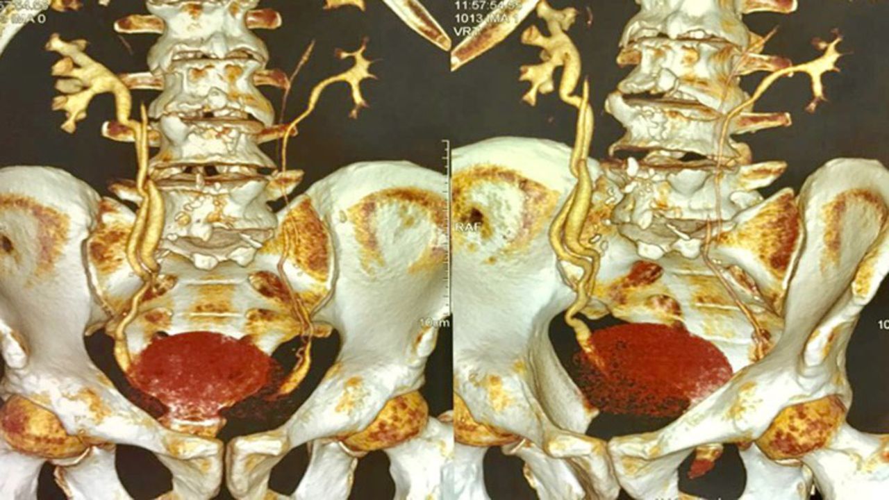
His complete blood haemogram and renal function tests were normal. On evaluation with ultrasound Kidney Ureter Bladder (KUB) and CT urography, there was presence of bilateral duplex kidneys with bilateral duplication of ureter with poorly functioning upper pole moiety of right duplex kidney with gross hydronephrosis and thinned out renal cortex with normal lower pole as shown in figure 1.
Three-dimensional reconstructed CT urography film showing bilateral duplex kidney with duplicated ureters with poorly functioning upper pole of right kidney.
Tc99m-diethylenetriaminepentacetate scan was done which confirmed non-functioning upper pole moiety of right duplex kidney with invariably hydronephrotic and thinned out parenchyma.
A laparoscopic transperitoneal heminephrectomy of non-functioning moiety of right duplex kidney was performed. After the induction of general anaesthesia, cystoscopy was carried out and a 6 Fr ureteric catheter was placed into the normal right lower ureter under fluoroscopic guidance after performing retrograde pyelography. Thereafter, the patient was positioned in a 45° lateral decubitus position and three ports were placed: 12 mm camera port at the umbilicus, a 10 mm port in the line joining the umbilicus and anterior superior iliac spine, and a third 5 or 10 mm port subcostally completing the triangulation.
After incising the white line of toldt and moving the colon medially, the duplicated ureters were identified medial to lower pole. The gerotas fascia was incised. The normal lower pole ureter was identified by the 6 Fr catheter that was previously placed. The diseased upper pole ureter was then dissected till non-functioning hilum was reached. To achieve such a meticulous dissection, we preferred using a combination of laparoscopic scissors and harmonic and staying as close as possible to serosa of the upper ureter.
Once the non-functioning renal hilum was identified, the renal artery and vein were clipped using haemolocks, two applied on each vessel. This made it easier to demarcate the upper pole non-functioning parenchyma from the functioning one. Using electrocautery and harmonic, we separated the upper pole from the rest of the renal parenchyma.
Duplex kidney is a rare entity with overall prevalence of 1/125 with 20% cases being bilateral and almost twice as common in females compared with males. It is usually associated with ureteroceles, vesicoureteral reflux and ectopic ureters accompanied by a poorly functioning upper pole segment.1
The embryonic ureter develops from the ureteral bud which itself develops as a branch from the mesonephric duct. As the bud grows and reaches the trigone, it gets absorbed leaving the ureteric orifice near its normal position. In case of ureteral duplication, the caudal ureter drains the lower pole and cephalic drains the upper pole. The upper pole is more susceptible to obstruction, and the lower pole has greater association with vesicoureteric reflux.2
Because of its manifestation like UTIs, urinary incontinence and voiding dysfunction during childhood diagnosis is made early and treatment is prompt with surgical intervention leading to resolution of symptoms in most cases.
Presentation in adulthood is different with flank pain and recurrent UTIs being more common.3
For management of duplex kidney, upper pole heminephrectomy with ureterectomy if the upper pole moiety is non-functioning or ureteropyelostomy/uretero ureterostomy with ureteric common sheath reimplantation if a functioning moiety is present via open approach is the standard of care.3
Our case was unique because we performed a laparoscopic transperitoneal heminephrectomy for managing such a rare condition.
Less than 100 such cases are reported in the literature.
Learning points
Bilateral duplex kidney with bilateral ureteral duplication is a rare anomaly with only a handful of reported cases, especially in adults.
Its manifestation during childhood includes urinary tract infections (UTIs), urinary incontinence and voiding dysfunction.
Presentation in adulthood is different with flank pain and recurrent UTIs being more common.
For management of duplex kidney, upper pole heminephrectomy with ureterectomy if the upper pole moiety is non-functioning or ureteropyelostomy/uretero ureterostomy with ureteric common sheath reimplantation if a functioning moiety is present is the standard of care.
Footnotes
Contributors SA: concept, design, supervision, processing, writing, manuscript and critical analysis. DS: concept and design. AS: supervision and processing. MS: concept and supervision.
Funding The authors have not declared a specific grant for this research from any funding agency in the public, commercial or not-for-profit sectors.
Competing interests None declared.
Patient consent Obtained.
Provenance and peer review Not commissioned; externally peer reviewed.