Article Text

Statistics from Altmetric.com
Description
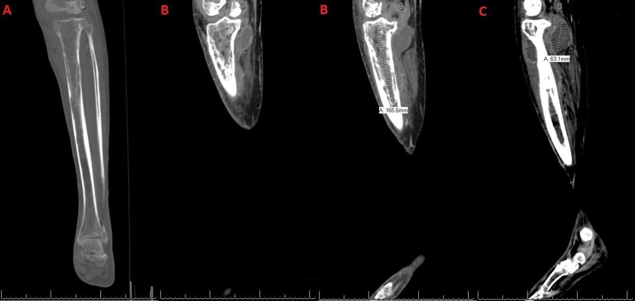
This is the case of a 43-year-old wheelchair-user man with a history of hepatitis C, cirrhosis, intravenous drug abuse and a 2-year history of chronic right lower extremity ulcers who presented to the emergency department with 4 days of worsening right leg pain. On physical exam, there was a new area of fluctuance on the anterior tibial surface. Labs included a white cell count of 9.3 K/µL, C reactive protein of 129 mg/L and erythrocyte sedimentation rate of 90 mm/hour. A CT scan demonstrated proximal tibia osteomyelitis with an intramedullary abscess, surrounding soft tissue abscesses and septic arthritis of the knee joint (figure 1).
(A) Permeative appearance of the proximal tibial metaphysis and diaphysis with areas of cortical lucency and endosteal scalloping, suspicious for osteomyelitis. (B) Intramedullary fluid collection within the proximal tibial metaphysis/epiphysis, containing hyperattenuating contents with two punctate foci of air measuring approximately 6.7×4.7×16.6 cm, suspicious for an intramedullary abscess. (C) Linear lucencies within the tibial cortex that communicate with multiple rim-enhancing fluid collections in the surrounding soft tissues surrounding the proximal and mid tibia, the largest of which measuring 3.8×5.0×6.3 cm.
The patient was taken urgently to the operating room for attempted limb salvage. Incision and drainage of the soft tissue abscesses was performed. A subsequent arthrotomy was performed with a large amount of pus expressed from the knee joint. Lastly, a large area of infected anterior tibial metaphysis was debrided, excised and saucerised.
Postoperatively, the patient was continued on intravenous vancomycin and cefepime. Intraoperative cultures grew Pseudomonas aeruginosa. Given the diffuse nature of the infection and the patient’s poor immune status, a multidisciplinary decision was made that amputation was the best treatment option. On hospital day 5, the patient underwent a right above knee amputation. Surgical pathology revealed chronic osteomyelitis.
Osteomyelitis is a severe, progressive infection of bone that can arise from a variety of mechanisms. Two commonly used classification systems include the Waldvogel and Cierny-Mader classifications.1–3
The more clinically useful Cierny-Mader classification is based on anatomy of the bone infection and physiological class of the host. In this system, there are four anatomic stages. In stage 1, the infection is confined to the medullary cavity. Stage 2 involves only cortical bone. Stage 3 involves both cortical and medullary bone, and the bone is stable without involving the entire diameter. Finally, stage 4 involves the entire thickness of the bone and is associated with loss of stability. The host is then classified: class A hosts have a normal immune system, class B hosts are immunocompromised and class C hosts are severely immunocompromised where treatment is considered worse than the disease.1–3
Our patient represents a challenging case of osteomyelitis given the acute intramedullary abscess and diffuse bone involvement. The bone had full thickness involvement, and the patient was biomechanically unstable on the affected leg. In addition, the host demonstrated systemic compromise. Based on the Cierny-Mader classification, this represents stage 4 osteomyelitis in a class B host. However, this classification system does not explicitly consider abscess formation.
Medical therapy is the mainstay of treatment for acute osteomyelitis, whereas the treatment for chronic osteomyelitis is primarily operative.1 Historically, treatment frequently required limb amputation. With advancements in surgical technique, the failure rate has been reduced to 10%–15%.2 Surgical management can be challenging as adequate debridement can leave large bony defects or dead space. Dead-space management is essential and may be achieved through complete wound closure, local tissue flaps or free flaps.3
Learning points
Chronic osteomyelitis is associated with significant morbidity and thus poses a challenge for both patients and physicians.
The management of chronic osteomyelitis requires an individualised approach that considers the dynamic nature of the infection as well as the host’s physiological, metabolic and immunological capabilities.
The Cierny-Mader classification is the most clinically relevant system used to describe osteomyelitis and is based on the anatomy of the bone infection and the physiological class of the host.
For long bone osteomyelitis, therapy includes adequate drainage, debridement, obliteration of dead space, wound protection and antimicrobials.
Newer surgical techniques that have improved limb salvage rates include Papineau bone grafting, Ilizarov external fixation, soft tissue muscle flaps, antibiotic impregnated beads and antibiotic impregnated bone grafts.
Footnotes
Contributors Conception and design, acquisition of data or analysis and interpretation of data: KB and SL; drafting the article or revising it critically for important intellectual content: all authors; final approval of the version published: all authors; agreement to be accountable for the article and to ensure that all questions regarding the accuracy or integrity of the article are investigated and resolved: all authors.
Funding The authors have not declared a specific grant for this research from any funding agency in the public, commercial or not-for-profit sectors.
Competing interests None declared.
Patient consent Obtained.
Provenance and peer review Not commissioned; externally peer reviewed.