Article Text

Statistics from Altmetric.com
Description
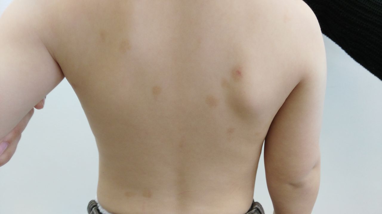
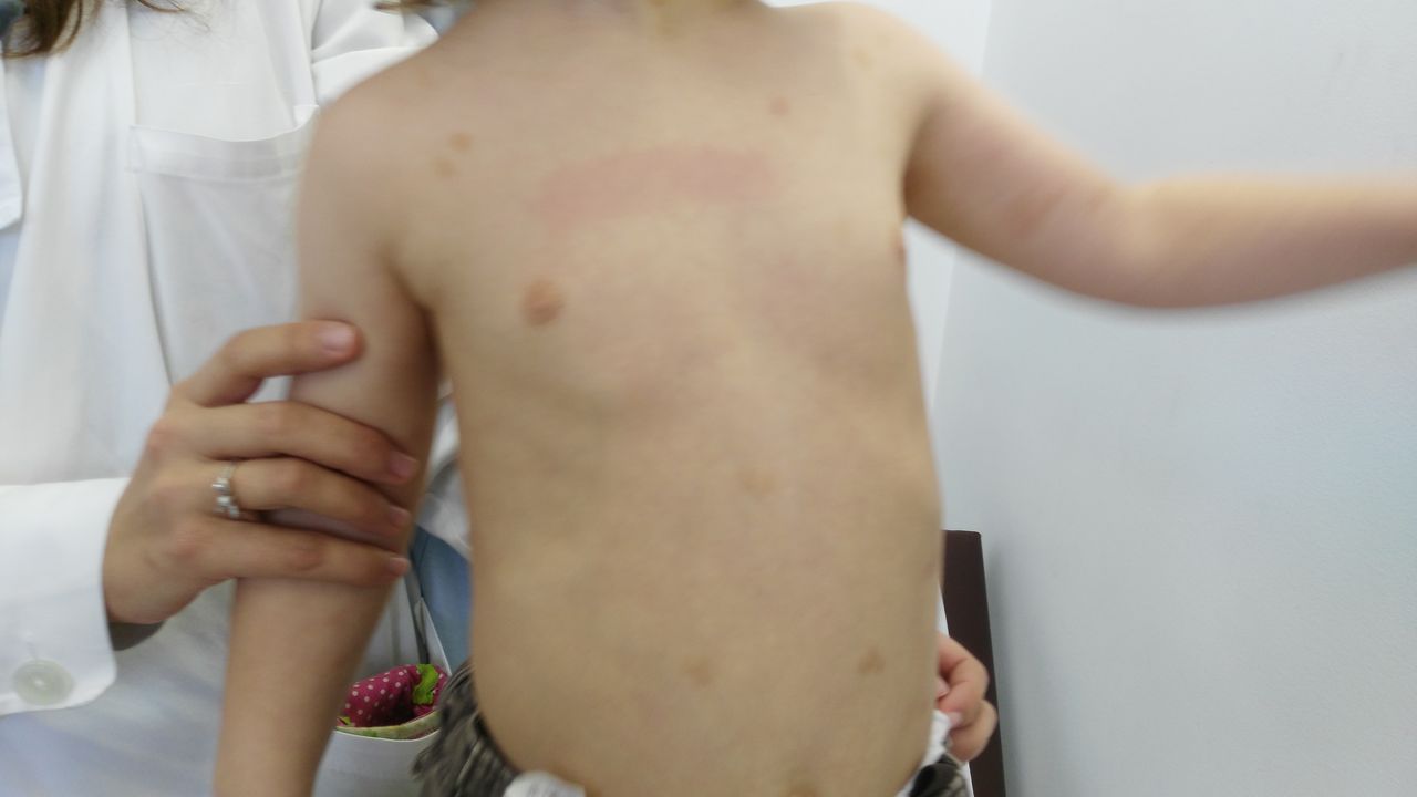
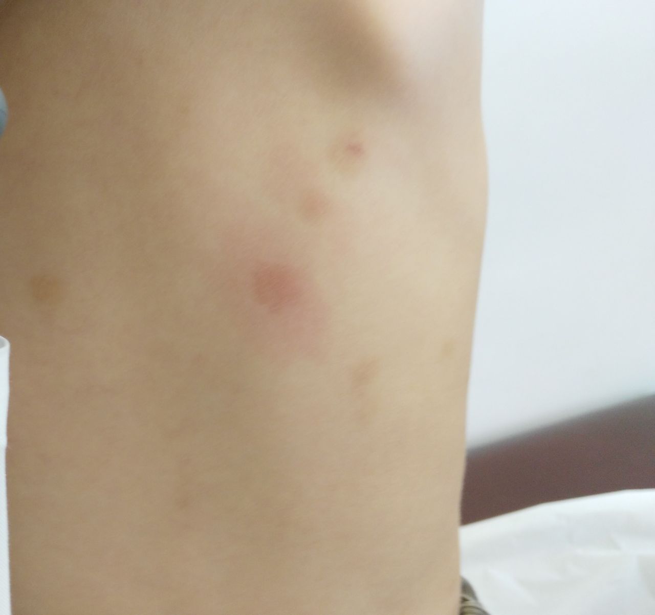
A 3-year-old boy presented to a paediatric consult with a rash consisting of reddish-brown non-pruritic spots. This rash initially appeared in his first months of age, with no identified triggers or associated symptoms and had been previously interpreted as eczema. Skin examination revealed irregularly bordered, hyperpigmented, cafe-au-lait macules on his trunk, neck and right forearm, the largest around 2 cm in diameter (figures 1 and 2). Rubbing one of the skin lesions elicited localised erythema—positive Darier’s sign (figure 3).

Skin lesions on the trunk (back).

Skin lesions on the trunk (front).

Darier’s sign.
Laboratory investigation showed both normal blood count and serum tryptase levels. He was diagnosed with urticaria pigmentosa (UP), a type of cutaneous mastocytosis. The patient is currently asymptomatic, without new skin lesions.
Mastocytosis is a rare and heterogeneous clinical entity, characterised by mast cell (MC) infiltration of tissues and organs.1 Based on the WHO’s classification, revised in 2016, mastocytosis can be divided into the following subgroups: cutaneous mastocytosis (CM), systemic variants (SM) and localised MC tumours. UP is the most common presentation of CM, diagnosed by characteristic skin lesions and supported by findings on skin biopsy.1 2
Our patient illustrates a case of UP in its classic paediatric form—hyperpigmented skin lesions appearing in the first 3–4 years of age and sparing the face, palms or soles.
However, when CM is suspected, SM must always be excluded. Children with SM may present with the same skin lesions, but have altered complete and differential blood count, organomegaly and sometimes inexplicable lymphadenopathy.1 3 Tryptase dosing is also important—a value of 20 ng/mL or greater is a minor diagnostic criterion for systemic disease, and an increased tryptase level is a risk factor for severe mediator release.2 3
Some syndromes characteristically have cafe-au-lait macules similar to those presented by this patient. The most common is neurofibromatosis type 1, an autosomal dominant genetic disorder caused by mutations in the NF1 gene. Its typical clinical manifestations are cafe-au-lait macules, axillary and/or inguinal freckling, Lisch nodules and neurofibromas. Juvenile xanthogranuloma could also be considered—a benign disease which classically starts to manifest in early childhood as solitary or multiple reddish to brownish skin papule(s), nodule(s) or plaque(s), most frequently on the head, neck or upper trunk.1 Other rarer syndromes to be considered are McCune-Albright, Legius and Noonan.1 A meticulous medical history and thorough physical examination, including Darier’s sign, are essential for the differential diagnosis.1 3
Despite having a good prognosis, CM can be associated with severe complications (such as anaphylactic shock) due to massive mastocyte degranulation. These episodes can be prompted by both emotional triggers and physical aggressors, such as drugs or certain types of food.1 3
Learning points
Searching for Darier’s sign is essential in the presence of typical brownish spots/macules in a child.
If cutaneous mastocytosis is suspected, serum tryptase is an important marker of systemic mastocytosis and/or severe mediator release.
Although urticaria pigmentosa has a good prognosis, parents should be informed of the potentially severe effects of massive mastocyte degranulation and its triggers.
Footnotes
Contributors All persons who meet authorship criteria are listed as authors, and all authors certify that they have participated sufficiently in the work to take public responsibility for the content. Identified and/or managed the case: HG, FN and SS. Idea for the article: SS and ARC. Performed the literature search: ARC. Wrote the article: ARC. Reviewed the article: HG, FN and SS. Guarantor: ARC.
Funding This research received no specific grant from any funding agency in the public, commercial or not-for-profit sectors.
Competing interests None declared.
Patient consent Not required.
Provenance and peer review Not commissioned; externally peer reviewed.