Article Text

Summary
Giant cell tumours (GCT) of the finger phalanges are extremely rare but have a high rate of recurrence. This report details the case of a GCT of the proximal phalanx of the fourth finger in a 64-year-old man. The patient was initially subjected to systemic neoadjuvant denosumab treatment, and subsequent aggressive curettage, sparing of the articular joints, local cryotherapy and autologous intercalary fibular bone graft. Finger function after surgery was considered satisfactory, despite limited proximal interphalangeal (PIP) joint motion. Aggressive local GCT recurrence was noted at the 32-month follow-up, with entire articular and diaphyseal phalangeal destruction. The patient refused amputation and, after analysing several reconstruction options, he was treated by entire en bloc resection and reconstruction employing a 3D-printed custom titanium implant. At the 24-month follow-up, the patient is free of disease and pain, and has a stable finger, good metacarpal–phalangeal joint motion, fusion of the PIP joint, a good Musculoskeletal Tumour Society score, and functional ability.
- oncology
- orthopaedics
- cancer intervention
This is an open access article distributed in accordance with the Creative Commons Attribution Non Commercial (CC BY-NC 4.0) license, which permits others to distribute, remix, adapt, build upon this work non-commercially, and license their derivative works on different terms, provided the original work is properly cited and the use is non-commercial. See: http://creativecommons.org/licenses/by-nc/4.0/
Statistics from Altmetric.com
Background
Giant cell tumour (GCT) is a benign, locally aggressive osseous neoplasm affecting the hand in 2% of cases.1 GCT of the phalanges are very rare and behave differently than GCTs found in more common locations (ie, the proximal femur); they also have very high recurrence rates.2–4 Here, we report the case of a patient who was initially treated according to the most recent guidelines for GCT local control: systemic neoadjuvant denosumab,5–7 aggressive curettage, local cryotherapy8 9 and reconstruction by autologous fibular bone graft. Although 80° proximal interphalangeal (PIP) joint stiffness existed , the graft fused, the spared articular joint was effective, and the overall result was satisfactory for most of the following 3 years. After GCT aggressive recurrence occurred 32 months postoperatively, and a finger amputation was considered. However, the patient, who had accepted the functional limitation, refused. While a second curettage was contraindicated, an en bloc resection was considered mandatory. The conventional surgical options were: arthrodesis with autologous graft, composite prosthesis (autograft or allograft bone diaphysis and prosthetic articular surface) and osteoarticular bone massive allograft. These surgical choices presented several functional or long-term concerns. For this reason, we used a custom 3D-printed titanium phalanx prosthesis to provide the patient with a long-lasting, stable and effective solution, although with analogous functional limitation. The construction of the implant was built based on measurements of the CT scans of the contralateral phalanx as a reference. The implant design included several proximal and distal holes for unabsorbable ligament reattachment.
Case presentation
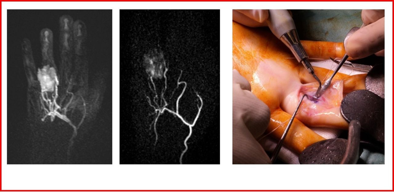
A 64-year-old male patient presented with pain and swelling at the level of the proximal phalanx of the fourth finger of his right hand, with limited motion of the PIP joint. The patient, a retired office worker, is right-handed and takes part in activities such as gardening, home improvement and bicycling. The diagnosis GCT of the proximal phalanx (figure 1) was histopathologically confirmed after incisional biopsy.

A 64-year-old male patient with a giant cell tumour at the fourth proximal phalanx.
To reduce aggressiveness and tumour vascularisation and spare the articular joint, the patient was treated with a 4-month preoperative course of systemic denosumab.5–7 After the 4-month treatment, he underwent surgery consisting of extensive curettage sparing of the articular surfaces, local cryotherapy8 9 and subsequent autologous fibular bone graft (figure 2).
First surgical treatment with neoadjuvant denosumab and cryotherapy followed by extensive curettage and autologous fibular bone graft.
The follow-up radiography at 3 months and 18 months after surgery showed a good outcome (figure 3) and satisfactory function.

Postoperative follow-up radiographies at 3 months and 18 months.
Thirty-two months after the surgical procedure, the patient presented an aggressive and destroying recurrence of the GCT at the same site involving the articular joint, which inhibited attempts at phalangeal sparing (figure 4).

Local giant cell tumour recurrence at 32-month follow-up.
The following treatment options were considered:
Amputation: this option was strictly refused by the patient. He declared that, despite 80° flexed PIP, the fourth finger was functional for precise manual dexterity.
Arthrodesis of the metacarpophalangeal (MCP) and PIP joints: since arthrodesis of MCP joint could have limited the patient’s leisure activities, it was not our treatment of choice.
Massive bone allograft of the first phalanx: this has been associated with a high risk of fatigue fracture with secondary loss of function.
Allograft prosthesis composite: because of the patient’s active lifestyle, this carried the same risk of fatigue fracture as massive bone allograft.
Autologous prosthesis composite: this was rejected because the patient had a previous donor site complication after fibular harvesting. Furthermore, silastic resurfacing offers encouraging long-term results in patients with low-demand activity level (eg, patients with rheumatoid arthritis), but these implants are at risk of failure and instability in active and high- demand patients.
For this reason, we decided to implant a titanium prosthesis, which can help maintain the range of motion (ROM) of the MCP joint and provide a stable PIP joint—even in flexion—with minimal risk of secondary fracture.
Treatment
We performed an entire first phalangeal resection and subsequent reconstruction using a prosthetic implant. A custom-made phalanx implant was created and shaped using CT scans of the contralateral finger as a reference. The implant design included proximal and distal holes for ligament attachments and reconstruction. The prosthesis was manufactured in a titanium alloy using electron beam melting technology and then coated with a layer of titanium nitride to allow for proximal and distal articulation with the surrounding bones (figure 5).

Complete phalanx resection and replacement with a 3D printed custom titanium nitride coated titanium implant. Proximal and distal ligaments have been reattached and reconstructed.
Outcome and follow-up
One year following the implant procedure, the ROM in the MCP joint was maintained, and the ROM in the PIP joint remained limited to 80° flexion (figure 6), as it was immediately after surgery. Two years following the implant procedure, the patient was disease free with a good Musculoskeletal Tumour Society score.10 The radiographs showed a fusion of the PIP joint, with dorsal subluxation, which may be related to difficulties in reconstructing the A2 and A3 pulleys. However, the joint remained stable and pain free, with subjective satisfactory results (figures 7 and 8).

Radiography at 1-year follow-up.
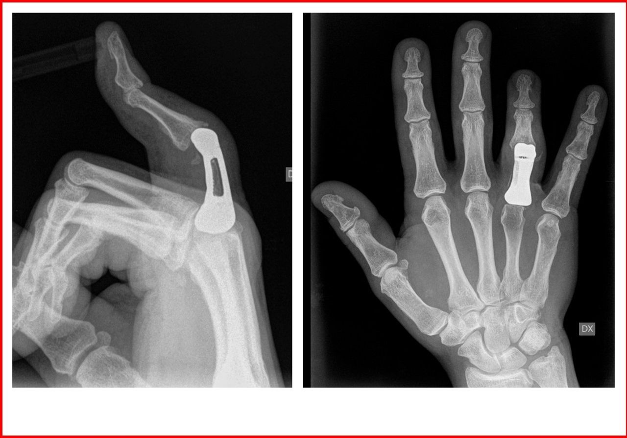
Radiography at 2-year follow-up. An intermediate dorsal subluxation and a fusion of the proximal interphalangeal joint is observed.

Functional outcome at 2-year follow-up. The joint is pain free and the patient has acceptable finger motion.
Discussion
This case represents the first such treatment of a GCT presentation in the phalanx, which is a rare presentation with a high recurrence rate. New metal 3D printing technologies allow for rapid manufacturing of customised implants tailored to the patient’s anatomy. In this case, after standard aggressive treatment failure, we implanted a custom titanium prosthetic. After 2 years, the patient has a stable and functioning MCP joint, a fused first interphalangeal joint and a good overall reconstruction; the patient is both pain and disease free. The patient has returned to all of his regular daily and leisure activities. We believe this surgical option was effective in this patient, after oncological failure of the previous, more conventional, treatment (ie, neoadjuvant denosumab, aggressive curettage, cryotherapy and autologous bone graft), with superimposable functional results.
After considering a range of surgical options (including amputation, arthrodesis, massive bone allograft, allograft prosthesis composite and autograft prosthesis composite), on the basis of our experience and with consideration of the functional demand of this patient, we decided to use a custom-made, 3D-printed phalanx prosthesis. The advantages of this approach are the mechanical resistance of the prosthesis and a potential permanent solution (although a longer follow-up period is needed to show this). Furthermore, the articular ROM was maintained according to preoperative measures, with complete MCP joint motion and rigid 80° flexion of the PIP joint. Because GCT of bone is a locally aggressive yet benign bone tumor, this prosthesis may represent a more acceptable option than amputation in many cases, even though it is more expensive.
For this reason, we routinely approach this disease by using curettage or resection and reconstruction. We are not aware of a published study that evaluates the outcome of primary amputation of a ray or limb for GCT of bone, except in cases of local or systemic complication (eg, infection).
We reserve amputation of the ray or finger for locally aggressive or recurrence of bone or soft-tissue sarcomas. For this reason, we accepted the patient’s firm refusal of finger amputation.
While further follow-up is needed, custom metal 3D-printed implants appear to be a good option for treatment of these and other oncological issues as a means of primary bone reconstruction or as a limb-salvage procedure after conventional surgical treatments are unsuccessful. However, some caution must be expressed with regard to the recovery of overall joint motion, with possible limitations in flexion as a final outcome.
Learning points
Giant cell tumours (GCT) of the finger phalanges are very uncommon, but they have a high recurrence rate.
We present a case of a GCT in the phalanx. A 3D-printed, custom-made 3D-printed phalanx implant was created and shaped according to the contralateral phalanx.
These implants can be a viable option for these and other oncological issues as a means of primary bone reconstruction after conventional surgical treatments have failed.
Footnotes
Contributors GB is the sole author of this case report.
Funding The author has not declared a specific grant for this research from any funding agency in the public, commercial or not-for-profit sectors.
Competing interests None declared.
Patient consent Obtained.
Provenance and peer review Not commissioned; externally peer reviewed.