Article Text

Statistics from Altmetric.com
Description
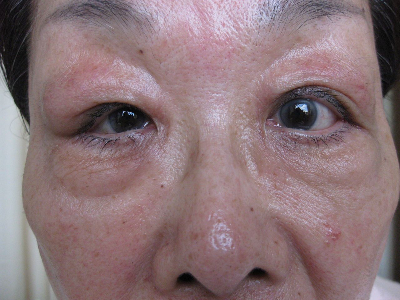
A 70-year-old woman suffered from bilateral lacrimal gland enlargement from 2 years ago (figure 1). Geranium-enhanced MRI disclosed diffuse enlargement of bilateral eyelids indicating IgG4-related disease (IgG4RD), especially Mikulicz’s disease. Laboratory tests showed elevation of serum IgG4 concentration and histopathological findings of the lacrimal glands showed IgG4-positive cell infiltration and obliterated veins by inflammatory cells, which consist of lymphocytes and plasma cells (obliterative phlebitis).

Bilateral lacrimal gland enlargement with predominance of right ones observed as a representative pathophysiological finding in this patient with IgG4-related disease.
Then, she was diagnosed with IgG4RD. We performed chest CT as screening for comorbidities of Mikulicz’s disease. Chest CT revealed left circumflex artery (LCX) wall thickening. Coronary CT showed thickening of the left anterior descending artery and LCX. For suppressing physiological myocardial uptake, the patient was asked to restrict carbohydrate intake 24 hours before 18F-fluorodeoxyglucose (FDG) PET/CT and to consume a low carbohydrate high fat protein permitted (LCHFPP) diet for lunch of the day before the examination, a low-carbohydrate diet the night before the examination and LCHFPP diet 4 hours prior to the examination.
FDG PET/CT showed uptake in the left ventricular anterior and lateral walls, and ascending aorta (figure 2A–D, I and K), indicating the comorbidity of IgG4-related periarteritis and subclinical pericarditis.

18F-fluorodeoxyglucose (FDG) PET/CT demonstrating the distribution of IgG4-related inflammation through active uptake of FDG in the left ventricular anterior wall (A), lateral wall (B) and the intermediate lesion of left ventricular myocardium and left circumflex artery wall (C, D). FDG uptake was confirmed to have disappeared by the follow-up study performed after the introduction of glucocorticoid therapy (E, F). FDG uptake was observed in the left ventricular wall, left circumflex artery wall (G) and ascending aorta (I). FDG uptake was confirmed to have disappeared in the left ventricular wall, left circumflex artery wall and ascending aorta by the follow-up study performed after the introduction of glucocorticoid therapy (H, J).
FDG uptake was also observed in lacrimal glands, retroperitoneum, pancreas and right common iliac periartery (figure 2M, O, Q and S). These findings suggested IgG4-related dacryoadenitis, retroperitoneal fibromatosis, pancreatic periarteritis and right common iliac periarteritis.
Glucocorticoid therapy was started from 30 mg/day of prednisolone. Bilateral lacrimal gland enlargement improved immediately after treatment. Serum IgG and IgG4 concentrations decreased from 2031 mg/dL and 785 mg/dL, to 758 mg/dL and 176 mg/dL 8 weeks later. C reactive protein (CRP) concentration also decreased from 1.06 mg/dL to 0.06 mg/dL. The uptake of FDG disappeared in the coronary artery and left ventricular wall, and ascending aorta (figure 2E, F, J and L). Coronary CT revealed decrease of coronary artery wall thickening of mid-LCX from 5 mm (figure 2G) to 3 mm (figure 2H) after the treatment.
Furthermore, FDG PET/CT suggested improvement of dacryoadenitis, retroperitoneal fibromatosis, pancreatic periarteritis and right common iliac periarteritis (figure 2N, P, R and T).
Then the dose of prednisolone was gradually tapered, and she is now being treated as an outpatient without recurrence of the disease. FDG PET/CT provides excellent information detecting systemic involvement in patients with IgG4RD. When the disease involved the cardiovascular system, it is particularly important to make an early diagnosis since it is recently reported that IgG4-related acute myocardial infarction and pericarditis resulted in death.1 While IgG4RD often responded dramatically to glucocorticoid therapy, sometimes the therapeutic effect was partial and glucocorticoid therapy could not avoid progression of the lesions.2 Then, metabolic response assessment of glucocorticoid therapy by FDG PET/CT was useful. FDG uptake in the IgG4-related lesions has been reported to have disappeared after steroid therapy.3 In this case, glucocorticoid therapy improved the size of the periarterial lesion and significant decrease in FDG uptake in parallel with decrease of serum IgG4 concentration. Thus, fusion images of PET/CT revealed the location of IgG4-related organs and was useful to evaluate the effect of glucocorticoid therapy for IgG4RD.
Learning points
This study showed that fusion images of PET and CT scans (PET/CT) helped identify IgG4-related organs and were useful to evaluate the efficacy of glucocorticoid therapy for IgG4-related disease.
18F-fluorodeoxyglucose uptake of the sclerotic lesions and periarteritis decreased in accordance with improvement of the clinical condition by glucocorticoid therapy.
A fusion imaging with the images of PET/CT was useful for identifying the location of its involvement and evaluating the effect of glucocorticoid therapy for IgG4-related disease.
Footnotes
Contributors JM wrote the manuscript with support from HT and WS. HT and WS supervised the findings of this work.
Funding The authors have not declared a specific grant for this research from any funding agency in the public, commercial or not-for-profit sectors.
Competing interests None declared.
Patient consent Obtained.
Provenance and peer review Not commissioned; externally peer reviewed.