Article Text

Statistics from Altmetric.com
- cardiovascular medicine
- emergency medicine
- respiratory medicine
- cardiothoracic surgery
- adult intensive care
Description
A 23-year-old Caucasian woman with a thin and tall body habitus presented to the emergency department with dizziness and chest pain. The pain had begun 12 hours before presentation without relation to exertion or trauma, radiating continuously to the neck and dorsum and being exacerbated by coughing or taking deep breaths.
The patient was previously healthy except for an episode of flu-like illness 2 weeks before presentation. She was not taking any medication and was a non-smoker. She had no relevant family history.
On clinical examination, she was conscious and reactive, afebrile and haemodynamically stable. She was eupnoeic and her oxygen saturation on pulse oximetry was 100%. Her breath sounds were normal on pulmonary auscultation, but the presence of a crunching sound synchronous with the heart beat was noted on cardiac auscultation (Hamman’s sign—video 1). A discrete subcutaneous emphysema was found on palpation of the left supraclavicular fossa. The rest of the physical examination was unremarkable.
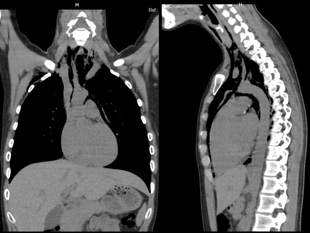
The total blood count, renal function, serum sodium and potassium, troponin I and C reactive protein were within normal range. The patient’s chest radiograph (figure 1) showed an abnormal radiolucent contour around the mediastinal structures, suggesting the presence of a pneumomediastinum. A thoracic CT (figure 2) excluded the presence of coexistent thoracic illnesses, such as structural lung disease or oesophageal perforation, and confirmed the diagnosis of spontaneous pneumomediastinum.
Hamman’s sign heard at cardiac auscultation.

Chest radiograph with an abnormal radiolucent contour around the mediastinal structures.

Thoracic CT showing the spontaneous pneumomediastinum.
After being assessed by the thoracic surgery team, the patient was admitted to the ward. She was treated conservatively with oxygen therapy, analgesia and avoidance of strenuous physical activity.
There was evidence of nearly complete reabsorption of the pneumomediastinum on the thoracic CT repeated 48 hours after admission. The patient was discharged home on the next day completely asymptomatic. She has been followed up in our outpatient clinic for 2 years without further events.
This case highlights the importance of a thorough physical examination in the assessment of chest pain. The presence of the Hamman’s sign on cardiac auscultation should prompt the consideration of the diagnosis of pneumomediastinum. Spontaneous pneumomediastinum is a rare entity that affects men more frequently than women.1 Its peak prevalence is seen in the second to fourth decades of life, especially among tall and thin patients.2 A chest radiograph may show signs of the presence of free air on the mediastinum, but the thoracic CT is essential to confirm the diagnosis and to exclude associated thoracic diseases such as gastrointestinal tract perforation or structural lung disease.3 Conservative treatment (oxygen, analgesia and avoidance of strenuous physical activity) is usually the preferred approach. The prognosis is typically excellent and the disease self-limited.1 2
Learning points
The presence of Hamman’s sign on physical examination should prompt the consideration of the diagnosis of pneumomediastinum.
Thoracic CT is essential to confirm the diagnosis of pneumomediastinum and to exclude concomitant thoracic pathology.
Conservative approach with oxygen, analgesia and avoidance of strenuous physical activity is the mainstay of treatment of spontaneous pneumomediastinum. The prognosis is excellent.
Footnotes
Contributors ARA wrote the body of the article and learning points. NFM and PR reviewed the whole article.
Funding The authors have not declared a specific grant for this research from any funding agency in the public, commercial or not-for-profit sectors.
Competing interests None declared.
Patient consent Obtained.
Provenance and peer review Not commissioned; externally peer reviewed.