Article Text

Summary
Sarcoidosis is a rare condition among native Saudis. It typically presents with asymptomatic chest radiographs, exertional breathlessness and cough. The coexistence of sarcoidosis and HIV is also rare, and the overlap of the symptoms makes their differential diagnosis challenging. Nevertheless, the outcome of sarcoidosis is favourable with or without the presence of HIV. We present a case of a 55-year-old native Saudi man with extremely atypical sarcoidosis presentation coexisting with HIV. This case highlights the association between the two pathologies, and the difficulties encountered in establishing a proper diagnosis in the presence of two overlapping diseases.
- general practice / family medicine
- immunology
- hiv / aids
- interstitial lung disease
This is an open access article distributed in accordance with the Creative Commons Attribution Non Commercial (CC BY-NC 4.0) license, which permits others to distribute, remix, adapt, build upon this work non-commercially, and license their derivative works on different terms, provided the original work is properly cited and the use is non-commercial. See: http://creativecommons.org/licenses/by-nc/4.0/
Statistics from Altmetric.com
Background
To the best of our knowledge, sarcoidosis has never been described as a cause of interstitial lung pathology associated with HIV in Saudi Arabia. In fact, it has rarely been associated with HIV in other countries and has been more commonly reported as a part of immune reconstitution inflammatory syndrome (IRIS) following highly active antiretroviral therapy (HAART).1 Sarcoidosis is a rare condition among native Saudis with the earliest mention in 1993 by Khan and colleagues where they reported 20 cases in their tertiary care centre.2
The epidemiological studies of sarcoidosis in Saudi Arabia or the Middle East are lacking, and therefore the incidence and prevalence of this condition are yet unknown. The most common symptoms of sarcoidosis in native Saudis are dyspnoea (48.64%), cough (44.59%), joint pains (39.18%), weight loss (28.37%) and fever (24.32%), as reported in the three studies conducted in Central, Western and Eastern Saudi Arabia in 1993, 1999 and 2011, respectively (table 1).2–4 Although a relative lack of cardiac, parotid, eye and central nervous system involvement has been observed in Saudi Arabia, the clinical presentation is comparable with that of the Western world.3 4 The most common stage of sarcoidosis observed in Saudi Arabia is stage II (51.35%) as opposed to stage I in the Western countries.2–4 There are no significant differences between the clinical presentation and pathology of sarcoidosis in patients with HIV infection and that of non-infected patients. Sarcoidosis manifestations are dependent on CD4+ cells, and the symptoms are more prominent with a CD4+ count >200 and subtle with CD4+ count <200.1 5 Therefore, sarcoidosis symptoms worsen with HAART as the CD4+ cell count raises following this therapy.5 Unfortunately, data regarding this correlation are lacking in Saudi Arabia and therefore cannot be compared with the West. This case report presents the first case of histologically proven sarcoidosis in Saudi Arabia and the Middle East that was diagnosed in a patient with low CD4+ HIV.
Case presentation
A 55-year-old native Saudi man was admitted with a 2-year history of weight loss and easy fatigability. The symptoms were associated with cyclic fever ranging between 37.4°C and 38°C every 2–3 days and a persistent cough producing yellowish sputum. The patient was diagnosed with hypothyroidism 3 years previously, smoked 60 packs of cigarettes a year and had been suffering from persistent swelling of the right neck and tongue pain for 9 years, and vague abdominal pain for 8 years. He had made multiple visits to the emergency department for these symptoms over the course of 2 years, but no apparent cause was identified even after extensive tests including abdominal ultrasound and contrast-enhanced CT of the neck, chest and abdomen done on November 2015—16 months prior to presentation—that was used for comparison. The ultrasound showed enlarged liver and haemangioma, and the contrast-enhanced CT scan of the neck and chest showed multiple bilateral neck lymphadenopathy and aortic arch atherosclerotic calcification, but no mediastinal lymphadenopathy or lung pathology. Enhanced abdominal CT was unremarkable. His blood work at that time showed no apparent abnormalities.
During examination, the patient was alert, conscious and oriented but was also distressed. He appeared ill and pale with temporal muscle wasting and generalised muscle weakness. His vital signs were stable, and he was breathing well. Cardiovascular examination was unremarkable, the chest was clear to auscultation with no adventitious sounds and abdominal examination revealed right upper quadrant fullness and mildly tender hepatosplenomegaly. The patient was admitted as a case of unexplained weight loss, possible chronic obstructive pulmonary disease (COPD) and hepatosplenomegaly in light of possible extrapulmonary tuberculosis (TB) for further evaluation.
Investigations
Ches X-ray (CXR) showed multiple small lung nodules, with the largest one in the lower right lung zone (figure 1). The blood analysis showed microcytic hypochromic anaemia with red cell distribution widht (RDW) of 21.6, and leucopenia with neutropenia and lymphopenia. Biochemical analysis showed blood ferritin levels of 613 µg/L, normal transferrin saturation, low serum iron, C reactive protein of 33.4 mg/L, amylase of 324 U/L, low normal calcium and erythrocyte sedimentation rate of 116 mm/hour (table 2). Abdominal ultrasound showed hepatosplenomegaly and redemonstration of haemangioma. CT scan of abdomen with contrast showed hepatosplenomegaly with multiple cystic lesions in the liver (figure 2). The findings were not conclusive, and we could not reach a satisfactory diagnosis. Therefore, after a lengthy discussion with the patient, we decided to explore other possibilities. At this point, the patient admitted to have risky sexual relationship outside marriage, something he had denied at admission. In light of this new information, we began an extensive serology work in addition to contrast-enhanced chest CT scan.
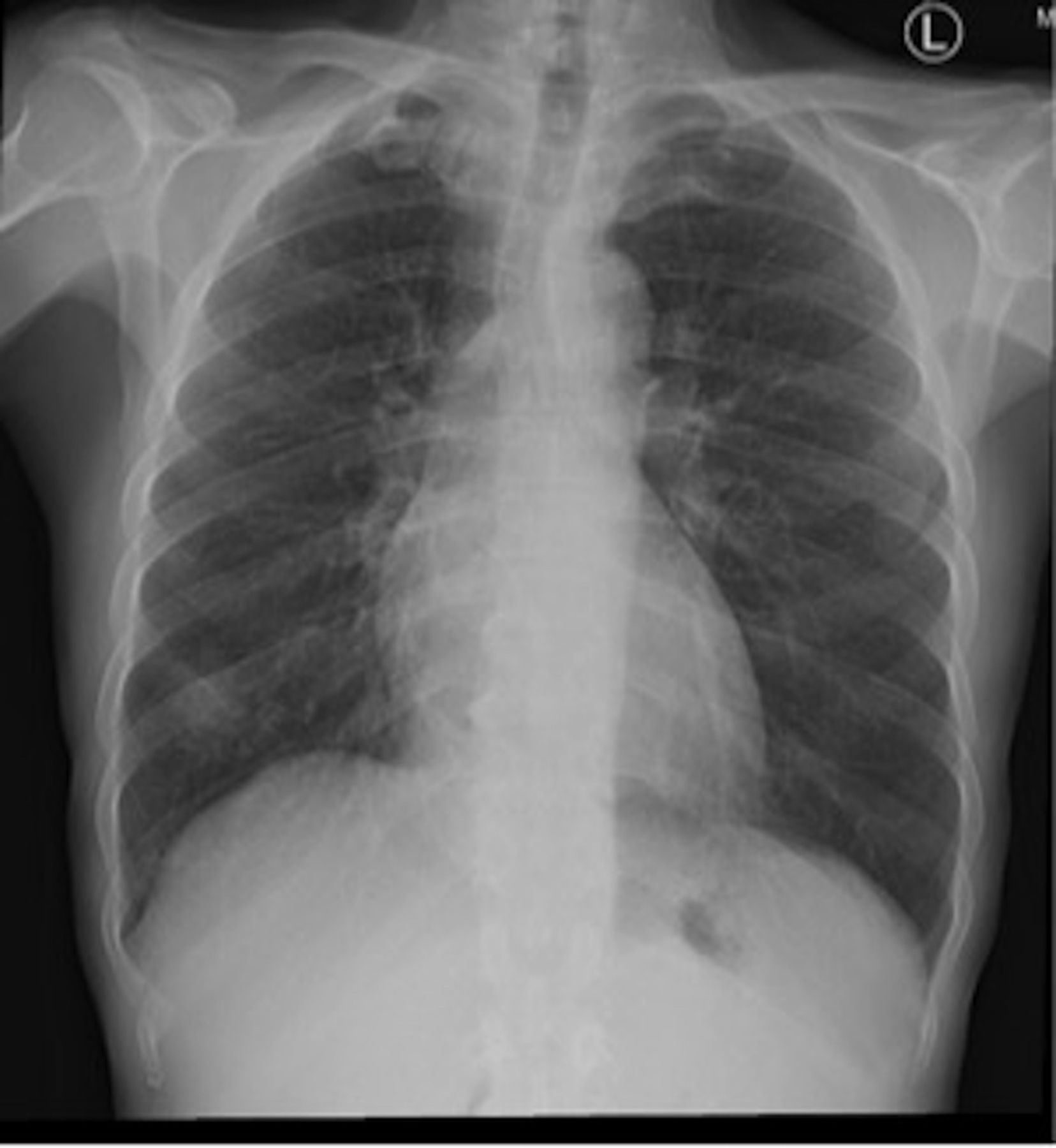
Chest X-ray taken at admission showing the right lung mass.
Patient blood work on admission
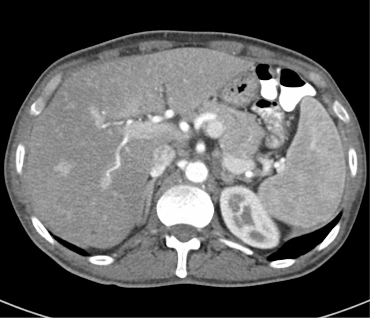
CT abdomen with contrast showing hepatosplenomegaly with multiple cystic lesions in the liver.
The chest scan showed significant interval developmental changes, multiple enlarged mediastinal lymph nodes, multiple pulmonary nodules mainly on the right side, with the largest one seen in the superior segments of the right lower lobe with speculated margin surrounding multiple satellite nodules, and redemonstration of previously seen subpleural cyst in the right lower lobe (figure 3). The differential diagnosis was broadened to include bronchogenic carcinoma owing to his high smoking index, and infectious diseases such as TB, Cytomegalovirus (CMV), HIV and HIV-related infection were also suspected. After gaining patient informed consent, CT-guided lung biopsy was obtained for definitive diagnosis while awaiting serology and sepsis screen results. The full sepsis screen and serology tests yielded negative results. However, CMV IgM tested positive, CMV IgG was negative and the HIV results were still pending. The biopsy showed chronic interstitial pneumonitis with non-necrotising granulomas. It was negative for acid-fast bacilli, fungi, atypia and malignancy (figure 4–6). The results at that time were highly suggestive of sarcoidosis. The findings were referred to cardiologists, ophthalmologists and rheumatologists to exclude sarcoid-related diseases. The patient was therefore started on a regimen of 40 mg oral prednisolone once daily. His condition started to improve, and he was discharged with instructions to come back the following week for his HIV and pulmonary function tests (PFTs) results, and for a further assessment. On follow-up, his PFTs showed restrictive lung pathology consistent with his diagnosis. The HIV test was positive, but his CD4+ count was low at 11 I/mm3. This was in contrast to published literature that has established the CD4+ dependency of symptomatic sarcoidosis in the presence of HIV, making this patient’s case extremely atypical and even more challenging.1 He then was referred to an infectious diseases specialist, and was started on HAART in addition to prednisolone. The symptoms of sarcoidosis did not worsen with the improvement of his CD4+ count over the next 6 months.

CT chest with contrast showing right lung granuloma.
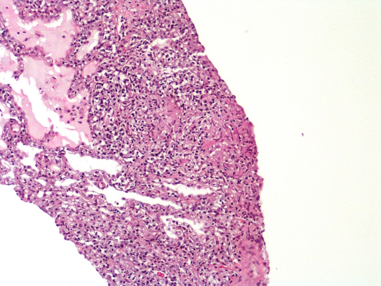
H&E staining of CT-guided right lung showing lung parenchyma with dilated alveoli, chronic inflammatory cell infiltrates and two non-caseating granulomas, ×100 magnified.

H&E staining of CT-guided right lung ×200 magnified, showing non-caseating granuloma in the interstitium with non-neoplastic lymphoid infiltrate.

H&E staining of CT-guided right lung showing two well-formed non-necrotising granulomas, ×200 magnified.
Differential diagnosis
TB (extrapulmonary): Although CXR did not show the typical features of TB, his clinical symptoms, in addition to the high TB incidence rate in Southern Saudi Arabia where the patient was domiciled, were highly indicative. However, lack of contact with any patients with known TB or family member/close associates with symptoms suggestive of TB, negative PPD testing, negative sputum culture and Acid fast bacilli (AFB) staining, lack of necrotising granuloma or AFB in the lung biopsy histopathology ultimately negated the diagnosis.6
Malignancies: The patient presented with a 2-year history of weight loss, easy fatigability, fever and expectoration. The temporal muscle wasting on examination, lung nodules on his CXR and a smoking history of 60 packs a year were suggestive of lung cancer. However, this diagnosis was excluded when the lung biopsy failed to show any atypical or malignant cells. Lymphomas and bowel malignancies were also suspected in light of the hepatosplenomegaly and scattered lymphadenopathy seen in imagining studies. However, his serum was negative for CA19–9, Carcinoembryonic Antigen (CEA), CA 125 and Prostate-specific antigen (PSA). Although the diagnostic value of the above is limited, that coupled with the negative biopsy made a diagnosis of cancer highly unlikely.
Undiagnosed COPD: Despite the patient’s smoking history and chronic coughs, and the nodular opacity seen in CXR, the pulmonology symptoms were unremarkable. Changes in CT chest scans occurring within less than 2 years and the restrictive pattern seen in his PFTs eventually excluded COPD as the main pathology.
Treatment
After establishing the diagnosis of sarcoidosis and exclusion of ocular, cardiac, neurological and joint sarcoid-related diseases, the patient was started on 40 mg oral prednisolone once daily for 2 months. Since he showed dramatic improvement within the first 2 weeks, the dose of prednisolone was gradually tapered over the following 3 weeks. However, due to his readmission with CMV retinitis, he was maintained on 15 mg orally once daily.
After the positive HIV test result, the patient was referred to an infectious diseases specialist and was started on HAART—200 mg emtricitabine/300 mg oral tenofovir disoproxil fumarate once daily and 50 mg oral dolutegravir once daily— in addition to prophylactic 960 mg oral co-trimoxazole once daily.
Right eye CMV retinitis was treated with 900 mg oral valganciclovir once daily for 3 months, along with intravitreal injections of 2 mg gancyclovir twice weekly (induction) which was lowered to 2 mg weekly with regular follow-up. He also received adjunct laser therapy.
Outcome and follow-up
The patient was readmitted after 17 weeks under ophthalmological services due to IRIS CMV retinitis. Unfortunately, despite treatment, he suffered subtotal macula-off retinal detachment and was referred to a specialist for retinal repair and vitrectomy, and the Out-patient Department (OPD) for follow-up after treatment.
Discussion
Sarcoidosis is a rare disorder which is diagnosed by exclusion, with an estimated incidence of 6 cases in 100 000 people per year in the USA.7 In Saudi Arabia, there have been only four major studies on sarcoidosis featuring approximately 180 cases. However, the incidence and the prevalence of sarcoidosis in Saudi Arabia have not been studied yet. The association of HIV and sarcoidosis, wherein patients with HIV infection typically show symptoms of sarcoidosis and other autoimmune diseases after initiation of HAART due to reactivation of the immune system, has been observed in several cases in the Western countries.5 Our patient was in his 50s, which is in keeping with sarcoidosis bimodal peak. His history of smoking, lack of dyspnoea and cough combined with lack of findings in chest examination were extremely atypical. The changes found in the plain chest X-ray were easily linked to undiagnosed COPD in light of his smoking history but were not as probative as his abdominal symptoms which were more alarming. Chronic sepsis, abdominal malignancy and extrapulmonary TB were on top of our differential diagnosis. However, the inconclusive septic screen, enhanced abdominal CT and PPD test results led us to explore other possibilities. Early scans of contrast-enhanced abdominal CT led us to pursue a chest CT, while fortunately we had a previous CT done 16 months ago to compare with. The developmental changes and enlarged lymph nodes found in the new contrast-enhanced chest CT encouraged us to carry out a lung biopsy to exclude malignancy since features of COPD were not found. Lung biopsy results showed non-caseating granuloma (figure 4–6). However, some of his symptoms were not conveniently explained by sarcoidosis (eg, pancytopenia, Hepatosplenomegly (HSM), non-granulomatous liver mass and cyclic fever) so HIV and other viral serology tests were performed. The result came out positive after the patient had been started on sarcoidosis therapy and discharged. Interestingly, his CD4+ count was low and according to available studies, sarcoidosis typically would not be observed with low CD4+ count due to its dependency on it.1 The patient’s CD4/CD8 ratio was also depressed, but this ratio has many limitations and is no longer used to diagnose sarcoidosis.8 His symptoms did not worsen after initiation of HAART and subsequent increase in CD4+ counts. The patient kept improving which may have been a result of the ongoing corticosteroid treatment.
Learning points
Sarcoidosis is still underdiagnosed and has not been properly studied in Saudi Arabia.
The association of sarcoidosis with HIV is quite rare, and the association between the two pathologies needs to be studied in Saudi Arabia.
There is a clinical overlap between the symptoms of sarcoidosis and HIV that makes their differential diagnosis quite challenging.
The outcome of sarcoidosis is similar in HIV-infected and non-infected patients.
Acknowledgments
Dr Rehab Bakr Fadag Consultant Histopathology MBChB, JBP, EBP, MIAC King Fahad Armed Forces Hospital, Jeddah, Saudi Arabia.
Footnotes
IAI and OME contributed equally.
Contributors OME and IAI were involved in study design and planning, data collection, data analysis and interpretation, preparation of the manuscript, literature analysis/search, and funding. HAA and MA were involved mainly in data collection and entry, literature analysis, English language revision and preparation of the manuscript.
Funding The authors have not declared a specific grant for this research from any funding agency in the public, commercial or not-for-profit sectors.
Competing interests None declared.
Patient consent Obtained.
Provenance and peer review Not commissioned; externally peer reviewed.