Article Text

Statistics from Altmetric.com
Description
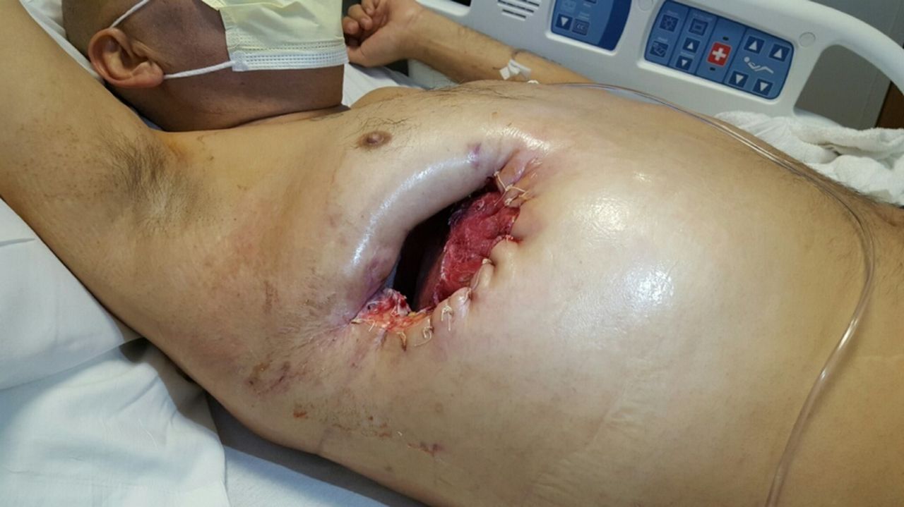
Coccidioidomycosis is a fungal infection by the Coccidioides genus and is usually caused by inhalation of the spores. Patients with diabetes are more likely to present with severe lung disease, especially cavitary lesions.1 2 We depict the case of a 47-year-old man with uncontrolled type 2 diabetes mellitus who presented with a right pulmonary lung abscess due to coccidioidomycosis and underwent a right thoracotomy with partial right upper lobe resection. Intraoperatively the pleura was found to be thickened, and the upper lobe had multiple adhesions and was perforated, creating a purulent bronchopleural fistula. He developed recurrent right-sided empyema due to the fistula and an attempt at a second thoracotomy was unsuccessful. After placement of three failed endobronchial valves, he ultimately had a tunnelled pleural catheter placed. Two weeks later the patient began draining frank pus from the site and a modified Eloesser flap (MEF) procedure was performed successfully (figure 1 and online supplementary video 1). He tolerated the procedure well and was discharged home with daily wound care. At 6-month follow-up, the patient had no recurrence of empyema. He unfortunately passed away later due to unrelated events. The MEF functions as a valve, permitting air to escape more rapidly than it enters, promoting negative pressure in the pleural cavity. This causes the affected lung to expand, sealing the inner opening of the flap and obliterating the empyema. MEF can permit bronchopleural fistulas to become bronchocutaneous, thus allowing adequate control of the infection.3
Right hemithorax status post-modified Eloesser flap procedure.
Right hemithorax status post-modified Eloesser flap procedure during inspiration and expiration.
Learning points
Patients with diabetes are more at risk to develop severe coccidioidomycosis and cavitary lung lesions.
Despite taking a back seat to antibiotic therapy and less invasive procedures, modified Eloesser flap still remains an option in the treatment of complicated recurrent empyema for patients who have exhausted other therapies.
Footnotes
Contributors OG: manuscript writing, images and video compilation. AI: manuscript writing, images and video compilation. AR: manuscript editing. YA: senior advisor, final manuscript approval.
Funding This research received no specific grants from any funding agency in the public, commercial or not-for-profit sectors.
Competing interests None declared.
Patient consent Obtained.
Provenance and peer review Not commissioned; externally peer reviewed.