Article Text

Summary
A 54-year-old man underwent decompressive craniectomy following a stroke. He further developed right lower limb ischaemia, and CT aortography revealed extensive aortic atherosclerotic disease. Urgent embolectomy prevented him from having a major amputation. He subsequently developed pulmonary embolism. This was initially treated with heparin followed by warfarin apart from antiplatelets and statin. A follow-up aortography at 3 months interval showed near complete resolution of atheromatous disease of the aorta. This report raises the possibility that apart from antiplatelets and lipid-lowering agents, anticoagulation may be responsible for resolution of such an extensive atheromatous disease and whether this can be considered as part of regular treatment.
- warfarin therapy
- vascular surgery
- stroke
This is an Open Access article distributed in accordance with the Creative Commons Attribution Non Commercial (CC BY-NC 4.0) license, which permits others to distribute, remix, adapt, build upon this work non-commercially, and license their derivative works on different terms, provided the original work is properly cited and the use is non-commercial. See: http://creativecommons.org/licenses/by-nc/4.0/
Statistics from Altmetric.com
Background
Severe atheromatous aortic disease with chronic smokers and other risk factors is known.
This leads to serious consequences, that is, stroke and peripheral vascular disease.
Various surgical and medical treatments have been used until now.
This case report raises a possibility that a combination therapy with statin, aspirin and warfarin may help resolve the atheromatous disease and prevent complications.
This may also obviate the need of potentially life-threatening surgeries.
Case presentation
A 54-year-old man with 40 years history of smoking was presented with a left-sided weakness and slurring of speech. On admission, CT brain scan showed extensive right middle cerebral artery territory infarct. He was started on aspirin. Two days later, his Glasgow Coma Scale (GCS) dropped to 9 (E2V2M5) and repeat CT brain scan done showed haemorrhagic conversion with significant mass effect, midline shift, along with the development of a hydrocephalus. He underwent decompressive craniectomy with intracranial pressure monitoring.
Ultrasound carotid revealed small plaques in the distal aspect of the right common carotid artery.
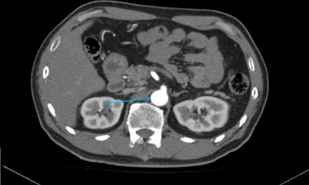
Three weeks after stroke, he developed acute left lower limb ischaemia. Urgent CT aortography (figure 1) showed severe and extensive atherosclerotic plaques (nearly 50% blockage) along with whole infrarenal aorta till its bifurcation, occlusion of the left popliteal artery from underlying thrombus, with partial occlusion of the left anterior and posterior tibial and peroneal arteries. A subsegmental pulmonary embolism in the right pulmonary artery was also detected during this scan.

Urgent CT aortography showed severe and extensive atherosclerotic plaques (nearly 50% blockage).
He underwent left popliteal embolectomy through popliteal approach on the same day, without any intervention of aortic athroma.
After careful discussion with the patient, family, neurologist, rehab physician and vascular team, postoperatively he received full anticoagulation (heparin followed by warfarin). Initially, he was treated with intravenous heparin with titration of dose based on activated partial thromboplastin time and careful monitoring of neurological status. A repeat brain scan was performed to ensure no deterioration or recurrence of bleeding. Further, he underwent multidisciplinary inpatient rehabilitation with partial functional recovery. At the time of discharge, he was wheelchair independent.
The medications during this period included: aspirin, warfarin and high-dose simvastatin. The repeat CT angiogram of aorta and left lower limb for postoperative follow-up after 3 months (figure 2). As compared with the previous CT angiogram, the eccentric mural thrombus narrowing the aorta showed near complete resolution along with restoration of the calibre of aorta, the left popliteal artery and the distal vessels. As per local anticoagulation guidelines warfarin was stopped at 3 months. Target international normalised ratio (INR) was also maintained at 2–3.
The repeat CT angiogram of aorta and left lower limb for postoperative follow-up after 3 months .
As the patient remained asymptomatic and did not have respiratory symptoms, further chest scan was not done. He has remained asymptomatic until now (7 years post-treatment).
Conclusion
In patients with embolic stroke, especially when carotid atherosclerosis has been established, possibility descending aortic atherosclerosis should be considered as this may help to prevent distal embolisation with possibility of limb loss especially in centre where vascular surgical access is not available.
Our single case report raises the possibility that apart from antiplatelets and lipid-lowering agents, combination therapy of statin and anticoagulation with warfarin may help complete resolution of atheromatous disease/aortic thrombus.
It also raises possibility if atherosclerotic disease of carotids may result in complete resolution with short-term anticoagulation, and future research on this treatment modality can be considered as a part of regular treatment for aortic thrombus.
Investigations
CT brain scan showed extensive right middle cerebral artery territory infarct.
Repeat CT brain scan done showed haemorrhagic conversion with significant mass effect, midline shift, along with development of a hydrocephalus.
Ultrasound carotid revealed small plaques in the distal aspect of the right common carotid artery.
Urgent CT aortography (figure 1) showed severe and extensive atherosclerotic plaques (nearly 50% blockage) along with whole infrarenal aorta till its bifurcation, occlusion of the left popliteal artery from underlying thrombus, with partial occlusion of the left anterior and posterior tibial and peroneal arteries. A subsegmental pulmonary embolism, the right pulmonary artery was also detected during this scan.
Treatment
He received warfarin for pulmonary embolism as per the local guidelines for 3 months duration, and dose was adjusted to keep the INR range within 2–3. He also continued aspirin (100 mg), statin (40 mg) during this period and as long-term treatment for his stroke.
Outcome and follow-up
He recovered physically to wheelchair independent state.
His repeat aortogram showed near complete recanalisation of descending thoracic aorta. During his 7 years of follow-up, he has remained asymptomatic from his pulmonary embolism point of view.
Discussion
Atheromatous aorta is only a part of systemic atherosclerotic disease. The risk factors for this are advancing age, hypertension and hypercholesterolaemia.1 Aortic atherosclerotic plaques are common in patients with coronary artery disease. These plaques may lead to embolisation into cerebral extremities or visceral circulation.1
Embolisation from plaques may be spontaneous or as a result of interventions, that is, coronary angiography or catheter studies and cardiovascular surgeries.1
Atherosclerotic plaques may lead to two types of emboli: thromboemboli and atheroemboli. Thromboemboli are common especially from thoracic plaques and they result from dislodgement of thrombus from atheromatous plaque, either from plaque rupture or force; these generally lodge in medium or large arteries. These results into transient ischaemic attack, stroke or acute limb ischaemia.1 Atheroemboli or cholesterol crystal emboli are under-reported probably due to their diverse presentations. As a result of disruption of atherosclerotic plaque, the debris of cholesterol crystals showers into circulation resulting in occlusion of arterioles that are <200 mm that typically affects organs.1
Cholesterol crystal embolism with atherosclerosis is significantly higher if trans-oesophageal echocardiography reveals protruding plaques, particularly more than 4 mm, and the presence of ulceration or superimposed mobile thrombi.1
The abdominal aorta and iliac arteries are commonly identified as source for lower extremity cholesterol crystal embolisation. In one surgical series of 62 patients, the aorta or iliac arteries were identified angiographically as the embolic source in 80% of patients.1
Atherosclerotic plaque in the femoral, popliteal or subclavian arteries may also be a source of extremity embolisation.1
Extensive atherosclerotic thrombus in non-aneurysmal aorta as reported in our patient is uncommon, and despite the availability of scanning techniques, literature revealed very few case reports on this subject.
Although aortic mural thrombus associated with aortic disease, such as aortic aneurysm and aortic dissection, is often seen, a thrombus in an apparently healthy aorta is not common; this is mostly as a result of aortic blood flow.2 3
The Virchow’s triad for thrombogenesis, featuring hypercoagulability, blood flow stasis and vessel wall injury, is known to be important in the thrombus formation, which is generally adopted for thrombosis of veins and small arteries.4 Cardiac arrhythmias are well known to cause thromboembolism, but not extensive thrombosis of aorta.2 3
Aortic mural thrombus in a non-aneurysmal, minimally atherosclerotic or normal aorta is uncommon cause of peripheral artery embolisation.5 6 This could be due to an under-diagnosed atheromatous disease, which may explain many cases of cryptogenic embolism.5
In our patient, the aorta appeared to be non-aneurysmal with such an extensive atherosclerosis leading to significant blockage. Literature search suggest that similar findings are more commonly associated with structural abnormalities of aorta.5 6
Treatment
In acute arterial thrombotic events, antiplatelet agents are used to prevent the incidence of arterial thrombosis and recurrent ischaemic events. But as coagulation is activated after plaque rupture, anticoagulation therapy is justified. Clinical evidence has shown that the combination of anticoagulant and antiplatelet therapy is more effective than either treatment alone.1 Statins reduce the rate of plaque development and size.3
Warfarin treatment may aggravate cholesterol embolism. It is concluded from other atherosclerotic manifestations that plaque-stabilising treatment with statin and ACE inhibitors is also beneficial.4
Apart from anticoagulation therapy, aortic surgery is alternative in patients who are fit for surgical intervention.5
A meta-analysis concluded that anticoagulation as first-line treatment for aortic mural thrombus is associated with higher risk of recurrence and complications and aortic surgery should be considered for patients who are good operative candidates and those patients who are at high risk of recurrence rates.5
Literature search revealed two cases of symptomatic thrombi of aortic arch treated with systemic tissue plasminogen activator, which had documented thrombus size, after follow-up for 6 months, showed free from thromboembolic events.7
In another single case study of 57-year-old man, without cardiovascular risk factors, thoracic CT scan showed pulmonary emboli and a thrombus of aortic isthmus. Trans-oesophageal echocardiogram after 49 days of warfarin therapy documented complete resolution of aortic thrombus.8
Our patient was also diagnosed with both pulmonary embolism and aortic atherosclerosis/thrombus without any evidence of aortic aneurysm.
He also was investigated for any underlying cause of cardiac arrhythmia, structural and functional abnormality of the heart. His 24 hours Holter was normal. Echocardiography suggested 60% ejection fraction, no motion wall abnormality and no evidence of left atrial or ventricular clot. This led us to conclude that the extensive thrombus in the aorta has developed from atherosclerotic disease, which may be related to his long-term smoking history.
He was treated with anticoagulation mainly in view of the pulmonary embolism. In the repeat CT angiograms, there was near total resolution of the thrombus along with atherosclerosis in the descending aorta. He did not have any complications from warfarin therapy and has been free of any further thromboembolic events for 6 years.
Learning points
Severe atheromatous disease of aorta may recanalise with combination therapy of warfarin, aspirin and statin.
Further trials on this subject may help establish treatment for carotid atheromatous disease.
In future, this triple therapy may be an alternative to more invasive procedures like carotid endarterectomy.
Footnotes
Contributors All the authors have contributed to the study. SDP contributed to the management and literature search and write up of the manuscript with FSH and VK. SK contributed to the initial management of the patient.
Funding The authors have not declared a specific grant for this research from any funding agency in the public, commercial or not-for-profit sectors.
Competing interests None declared.
Patient consent Obtained.
Provenance and peer review Not commissioned; externally peer reviewed.