Article Text

Statistics from Altmetric.com
Description
Rhinophyma meaning ‘nose growth’ in Greek is a relatively common condition that describes thickening of the nasal skin with enlargement of the sebaceous glands. While not fully understood, it is believed to be a result of vascular instability causing leakage of fluid into the tissues. This subsequently triggers inflammation and scarring.1 Treatment is initially medical; systemic isotretinoin has been shown to reduce the bulk of rhinophyma. Many surgical techniques have also been described, all of which involve tissue removal. Previous literature has demonstrated other skin conditions mimicking this diagnosis including angiosarcoma, squamous cell carcinoma and sarcoidosis.2
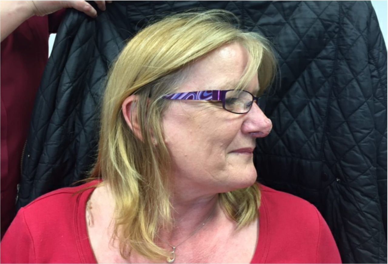
A 52-year-old woman was referred to ear, nose and throat (ENT) from dermatology for surgical management of rhinophyma. She described a 3-year history of an increasing swelling on the tip of her nose (figure 1). During this time, she underwent extensive conservative treatment with dermatology including a course of isotretinoin to little effect. The appearance of her nose had been causing a significant influence on her mental health and social well-being. She had been prescribed antidepressants by her general practitioner and was reluctant to progress in her career, as this would include more contact with the general public.

Clinical features consistent with rhinophyma centred on the tip of the nose, giving a markedly distorted appearance to the nasal tip.
At first skin incision with a scalpel, instead of the anticipated rhinophyma, a large subdermal mass was unexpectedly encountered (figure 2). This was removed without difficulty, and a good cosmetic result was achieved by reshaping/regrafting the removed skin flap. The mass was sent for histology which surprisingly identified it as a schwannoma. Follow-up at 2 weeks and 3 months confirmed a good cosmetic outcome and patient satisfaction (figure 3). No further management was required.

Intraoperative image displaying rubbery soft tissue mass delivered through initial incision.

Appearance at 3 months following surgery.
Schwannomas are benign tumours of the nerve sheath that are composed of Schwann cells. They account for between 25% and 45% of head and neck tumours of which 4% are found in the nasal cavity. Schwannomas of the nasal tip are rare with only a handful of cases previously being reported.3 Preoperative diagnosis can be made based on biopsy or imaging studies where there is clinical suspicion. This was not undertaken in this case as both ENT and dermatology agreed on the clinical diagnosis of rhinophyma.
Preoperative MRI may have helped identify the schwannoma given the tumour’s predictable signal characteristics. A number of signs on MRI have been described including the split-fat sign, target sign and fascicular sign.4 Histology typically shows an encapsulated well-circumscribed lesion beneath the uninterrupted epidermis. The tumour is composed of areas that are composed of different cellular densities.
Learning points
Nasal tip schwannoma is a rare differential diagnosis when considering rhinophyma.
Biopsy and imaging prior to surgery may help where there is diagnostic uncertainty, therefore allowing better surgical planning.
If this patient had undergone treatment by microdebridement, a rapid and good cosmetic outcome may have been more difficult to achieve.
Footnotes
Contributors TG wrote the manuscript. AHH, JM and SM were involved in clinical care and revision of manuscript.
Competing interests None declared.
Patient consent Obtained.
Provenance and peer review Not commissioned; externally peer reviewed.