Article Text

Statistics from Altmetric.com
Description
An 85-year-old woman presented to the ear, nose and throat clinic with a 2-week history of left-sided otorrhoea and pruritus of the ear. Examination of the left external auditory canal (EAC) revealed a polypoidal lesion and purulent discharge (figure 1). The tympanic membrane was intact. The suspicious lesion prompted imaging, including CT neck and thorax (figure 2). An ultrasound scan of the parotid and neck showed no metastatic disease. A biopsy was undertaken and histology demonstrated a basal cell carcinoma (BCC). The patient was managed with a staged procedure. Stage 1 consisted of a wide local excision of the BCC, with a 4 mm margin. Frozen section was not available; therefore, a second stage was needed to achieve clear margins using a sleeve resection. Fortunately, the disease was limited to the cartilaginous ear canal, hence did not require further resection or reconstruction. This management approach was successful and the patient remains in follow-up.
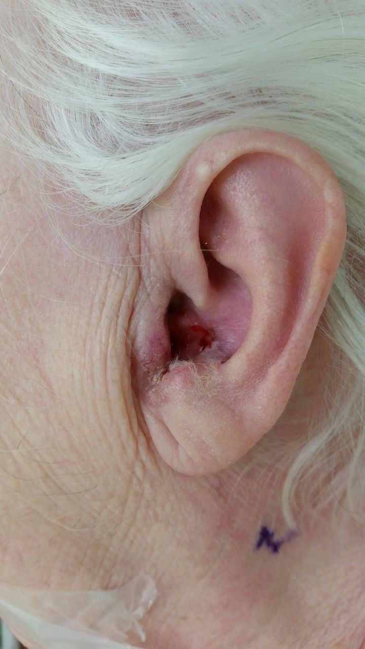
The innocuous appearance of a basal cell carcinoma in the external auditory canal, highlighting the difficulty in making a correct diagnosis.

CT head and neck scan illustrating the cutaneous lesion involving the left pinna and external auditory canal. This shows the extent of disease and how intracranial invasion can occur in advanced disease. (A) Axial view. (B) Coronal view.
The annual incidence of EAC carcinomas is around one per million.1 However, BCCs are known to be locally aggressive, which can be terminal if intracranial invasion occurs.2 Bony canal involvement may require temporal bone resection, and extensive lesions may need pinna removal and reconstruction. Early and correct diagnosis is therefore essential, specifically by healthcare professionals who frequently manage skin conditions. Out of all primary care consultations in England and Wales, 24% are related to skin conditions according to published data.3 Greater awareness of BCCs occurring in the EAC will lead to earlier diagnosis and ultimately improve outcomes for this disease.
Learning points
Basal cell carcinomas occurring in common sites such as pinna carry a good prognosis. Basal cell carcinomas of the external auditory canal can be terminal.
Healthcare professionals in primary and secondary healthcare should be aware of this basal cell carcinoma presentation to allow for early diagnosis and management.
It should be explained to patients with suspected external auditory canal lesions the necessity of early treatments and surgery to avoid large defects requiring reconstruction.
Footnotes
Contributors All authors participated in the work as a whole. The manuscript was written by MK, with support from MD and SK on the whole project.
Competing interests None declared.
Patient consent Obtained.
Provenance and peer review Not commissioned; externally peer reviewed.