Article Text

Statistics from Altmetric.com
Description
Pyoderma gangrenosum (PG) is defined as a neutrophilic dermatosis, not related to any infections or gangrenous causes.1 It usually appears with an underlying systemic disease. We report a 55-year-old man who presented with painful bilateral skin lesions on legs with no associated constitutional symptoms. Patient had a history of untreated hepatitis C infection with negative cryoglobulinaemia screen. Surgical debridement was performed in the emergency department, at that time without a diagnosis and a biopsy plus culture of the skin was done which disclosed no evidence of microorganism, vasculitis findings and perivascular lymphoplasmacytic infiltrate. A repeat biopsy showed neutrophilic infiltrate. Clinical and histopathological diagnosis of PG was made and patient was started on intravenous steroids with some improvement,2 followed with clinical deterioration that prompted intravenous immunoglobulin therapy with excellent results.3 Patient continues to get weekly wound care and was being seen by infectious disease and rheumatology service. The importance of these images is to remind us that clinical acumen supersedes ancillary tests, since the initial biopsy was only suggestive, and the diagnosis of PG was made based on clinical criteria with combination therapy being adequately started using intravenous steroids, intravenous immunoglobulin and weekly wound care with excellent results (figures 1 and 2).
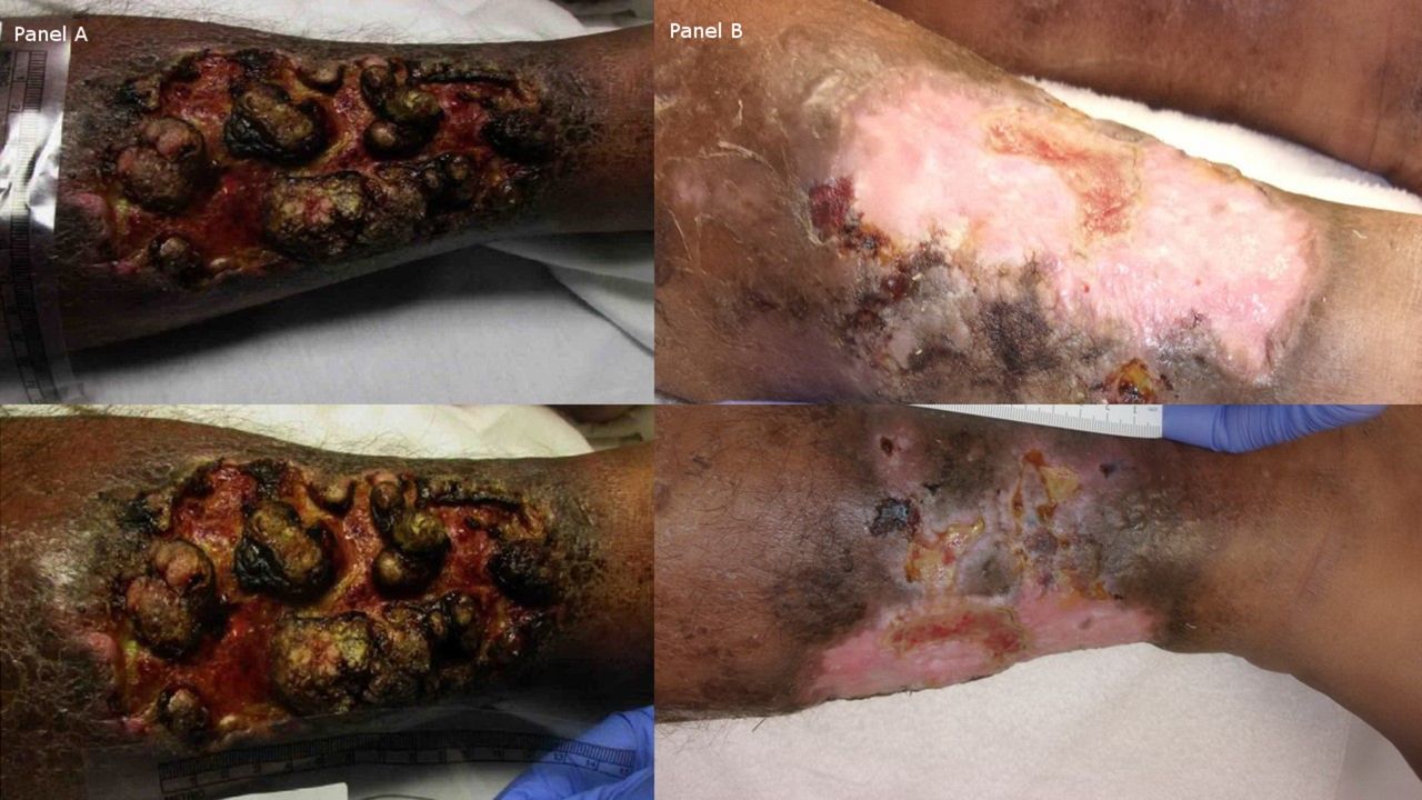
Panel A shows the right lower extremity before treatment, pustular base with hyperkeratotic lesions. Panel B shows right lower extremity after treatment, clear base and resolution of the keratotic lesions.

Panel A shows the left lower extremity before treatment, pustular base with hyperkeratotic lesions and crusting, with some areas of haemorrhages. Panel B shows left lower extremity after treatment, clear base and resolution of the keratotic lesions. Upper image of the panel B did show some pustular lesions.
Learning points
The diagnosis of pyoderma gangrenosum (PG) has to be based in a combination of both clinical and histopathological features, clinical diagnosis can be made based on suspicions, like in our case where although biopsy was only suggestive of PG and only subsequent biopsy showed the typical neutrophilic infiltrate.
Treatment should be multidisciplinary, as exemplified by this case managed with intravenous steroids, intravenous immunoglobulin and weekly wound care.
PG is a rare condition and can sometimes present as a long-standing non-healing ulcers refractory to antibiotic treatment. Awareness is needed in the medical community, so is always present in our differential diagnosis.
Footnotes
Contributors AI was responsible for image composition, manuscript writing and editing. OG, YA and KB were involved in the manuscript editing. YA and KB were responsible for the final manuscript approval.
Competing interests None declared.
Patient consent Obtained.
Provenance and peer review Not commissioned; externally peer reviewed.