Article Text

Statistics from Altmetric.com
Description
A 12-month-old boy presented with a history of respiratory distress and stridor since the first month of life. He had been repeatedly misdiagnosed as recurrent croup. The stridor was biphasic, with a more pronounced inspiratory component, and was exacerbated by agitation and supine positioning. He had a large posterior cervical haemangioma, whose extension had never been studied by imaging methods, and delayed growth. Flexible bronchoscopy showed a multilobulated subglottic haemangioma (SGH), occupying more than 70% of the tracheal lumen. MRI showed an angiomatous malformation with the epicentre at the hind head that extended inferiorly to the cervical planes, reaching the median line in retropharyngeal planes with inferior extension (figures 1 and 2). He underwent systemic and intralesional tracheal steroids injections with partial improvement and laser therapy at 5 years of age. Today he is an asymptomatic 14-year-old adolescent with normal growth and development.
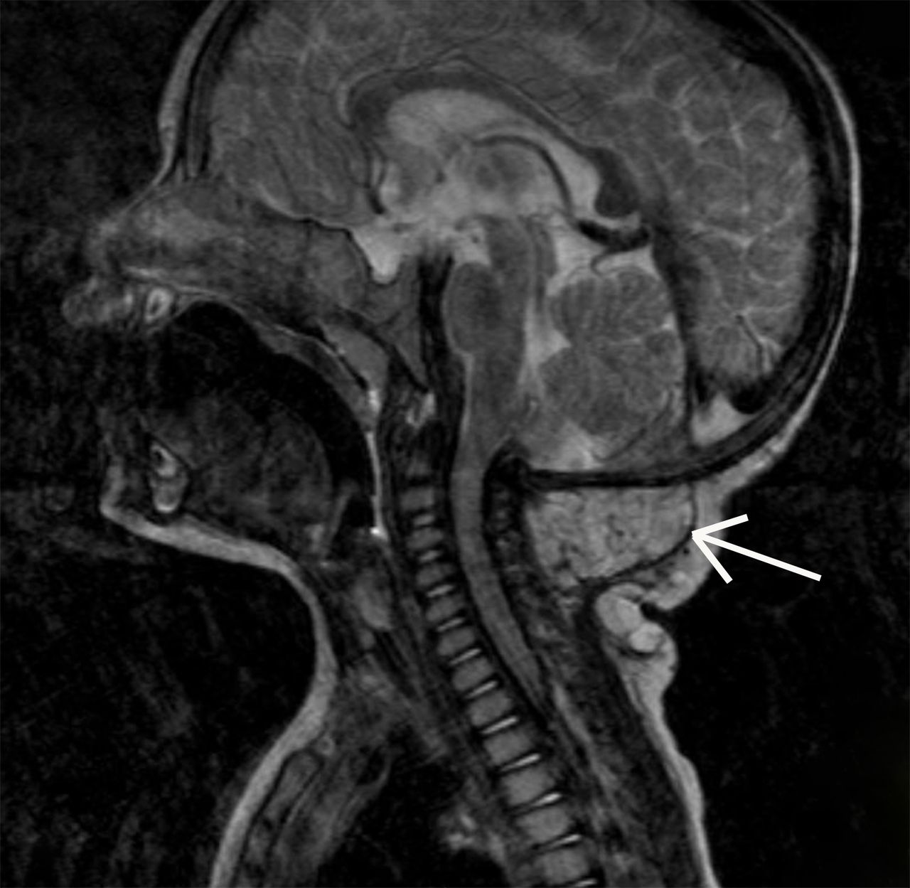
Cervical MRI examination, sagittal plane, showing extracranial angiomatous formation with epicentre in the posterior cervical region (white arrow).

Cervical MRI examination, axial plane, showing multisegmented angiomatous formation that penetrates the different cervical spaces.
Infantile haemangiomas are the most common vascular tumours of childhood, affecting 5% of all infants.1 SGH represents 1.5% of the congenital laryngeal abnormalities. Almost 50% of them are associated with cutaneous haemangiomas in the face.2 There is a correlation between the localisation of the cutaneous haemangioma and the risk of SGH (chin, lip, neck, preauricular region).2 Their rapid growth may lead to airway obstruction.2 Currently, when medication is warranted for infantile haemangioma, oral propranolol is the drug of first choice.3
Learning points
The presence of cutaneous large haemangioma in the chin, lip, neck, preauricular region along with stridor/respiratory distress, must raise a high suspicion of multilobulated subglottic haemangioma.
Airway haemangiomas represent a potentially fatal complication of infantile haemangiomas.
Oral propranolol is now the first-line treatment.
Acknowledgments
none
Footnotes
Original reference: none
Contributors JCO: Follow-up of the patient, discussion of conduct, conception and design, acquisition of data, literature research and conception of the paper. IA: patient's diagnosis and treatment, literature research and revised the final version of the article. AG: patient’s diagnosis and treatment, literature research and revised the final version of the article. CM: patient’s diagnosis and treatment, follow-up of the patient, literature research and revised the final version of the article.
Competing interests None declared.
Patient consent Guardian consent obtained.
Provenance and peer review Not commissioned; externally peer reviewed.