Article Text

Statistics from Altmetric.com
Description
A middle-aged woman presented with a 12-hour history of generalised abdominal pain and septic. Her medical history included chronic alcoholism and a 40 pack-year smoking history. Laboratory tests revealed raised inflammatory markers and a macrocytosis. There were no liver, renal or clotting derangements, and haemoglobin and platelet levels were within normal limits. Blood gas analysis showed acidaemia and raised lactate. A CT scan of the abdomen revealed pneumoperitoneum, extensive portal venous gas and pneumatosis intestinalis throughout the entire imaged gut—from distal oesophagus all the way to anus—suggestive of complete intestinal necrosis (figures 1–3). The mesenteric arteries appeared patent. The patient rapidly deteriorated and died within 3 hours of admission.

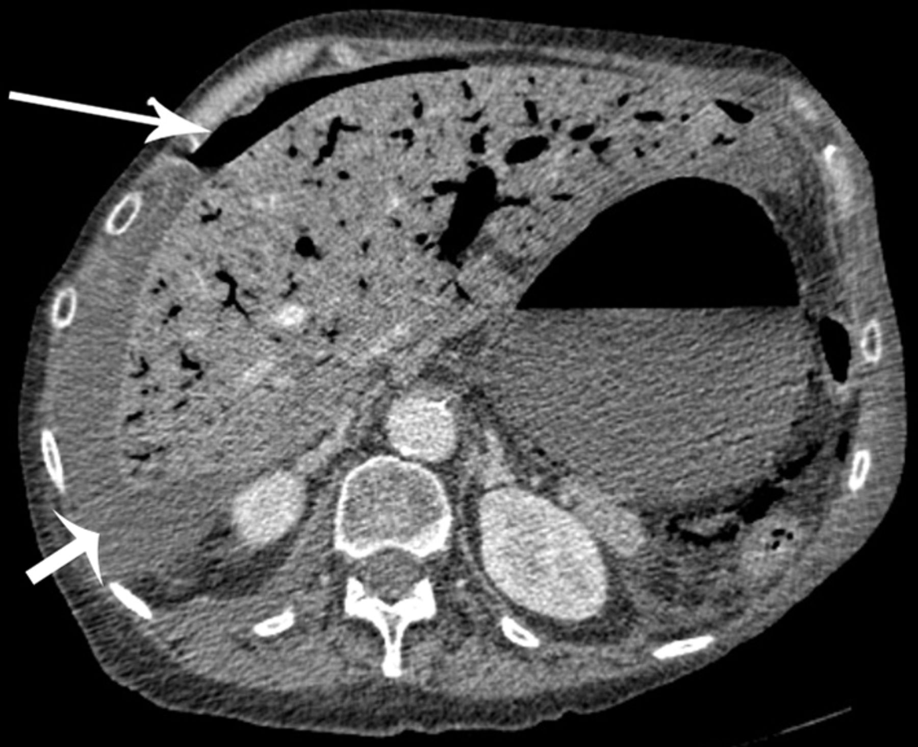
Axial CT image of upper abdomen showing extensive portal gas, pneumoperitoneum (long arrow) and intra-abdominal free fluid (short arrow). Gastric wall intramural gas (pneumatosis) is visible.

Axial CT image through pelvis showing significant intramural gas throughout the bowel loops (arrow).

Coronal CT image demonstrating intramural gas in the oesophagus (long arrow). This is also present in the stomach, small and large bowel, including bowel in the pelvis (short arrow).
Such CT appearances are in keeping with rare interlinked conditions adult necrotising enterocolitis and non-occlusive mesenteric ischaemia; however, such extensive necrosis of the entire intra-abdominal bowel has not been reported previously. Previous studies have hypothesised either a primary infective cause or a hypoxia-reperfusion injury from a primary vascular event leading to secondary infection from bacterial translocation across the injured bowel wall.1 Despite the unclear aetiology, a previous review of four cases describes common risk factors—chronic alcoholism and smoking—also present in this case.2
This case describes necrosis of the entire bowel with extensive portal venous gas and pneumoperitoneum—clearly incompatible with survival. Surgical options are limited. These appearances should be considered a preterminal sign and a high index of suspicion for intestinal necrosis must be considered in alcoholics and smokers who present with an acute abdomen.
Learning points
Extensive necrosis of the bowel with portal venous gas is usually considered a preterminal sign—surgical options are limited at this stage.
A high index of suspicion for intestinal necrosis must be considered in alcoholics and smokers who present with an acute abdomen.
Footnotes
Contributors MMcK: conception of the article, data collection, drafting the article and final approval of the version to be published. DC: critical revision of the article and final approval of the version to be published.
Competing interests None declared.
Patient consent Detail has been removed from this case description to ensure anonymity. The editors and reviewers have seen the detailed information available and are satisfied that the information backs up the case the authors are making.
Provenance and peer review Not commissioned; externally peer reviewed.