Article Text

Statistics from Altmetric.com
Description
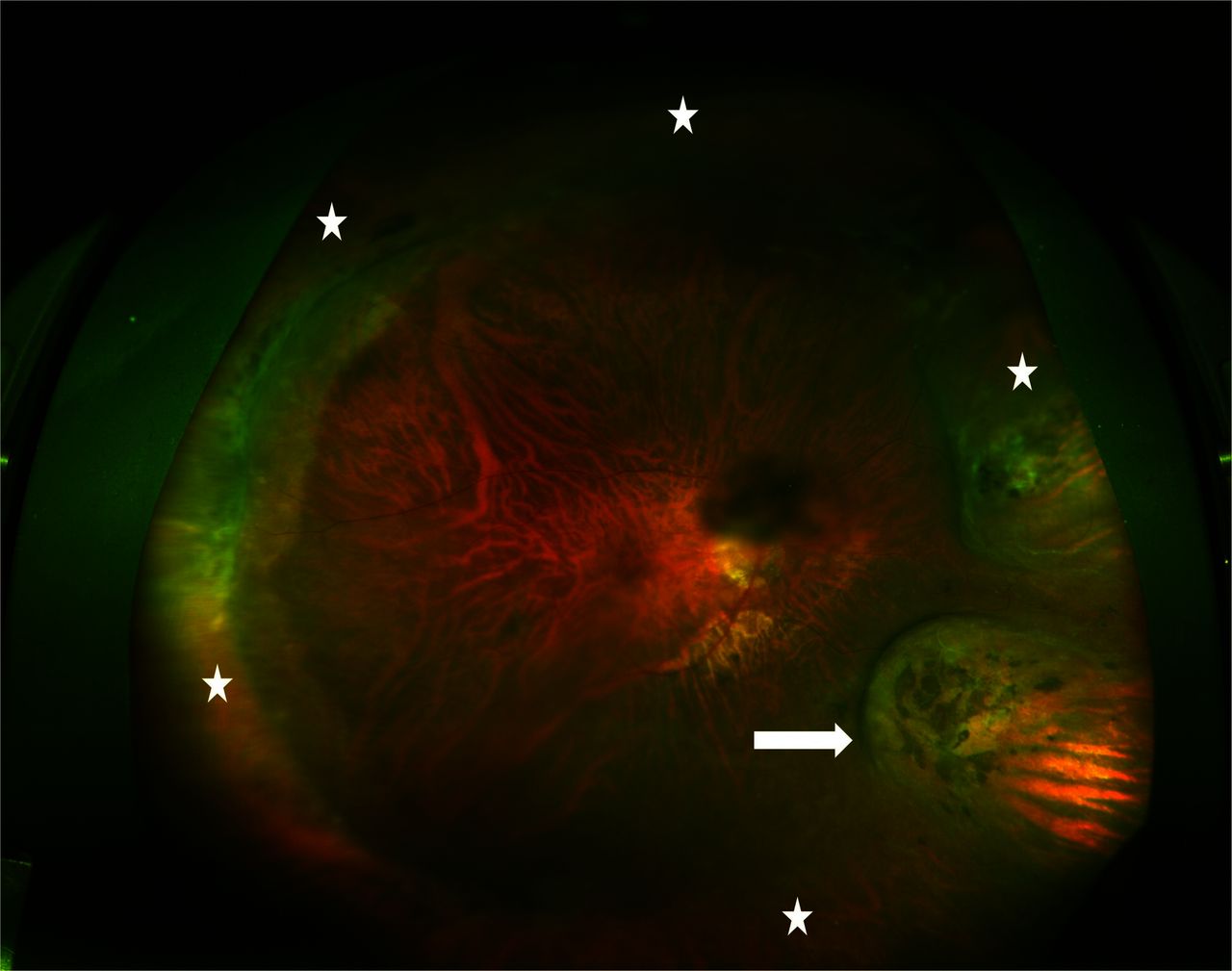
A 78-year-old male presented with blurring of vision in right eye (RE) since 2 months. He was diagnosed to have retinal detachment in RE 27 years ago, which had been successfully repaired with scleral buckling surgery. Retinal holes had been treated with laser prophylaxis in left eye (LE). On examination, visual acuity was 6/24 in RE and 6/9 in LE. Both eyes had posterior subcapsular cataract (RE>LE). The retina was attached in RE, while features of high myopia, like fundus tessellation and peripapillary crescent, were evident. Indent of a radial explant was noticeable in the inferior nasal quadrant along with indent of an encirclage band (figure 1). Lattice degeneration and adequately lasered retinal lesions were seen in both eyes. The posteriorly placed radial explant was noted as a raised and hyperechoic structure with corresponding acoustic shadow on ultrasound B scan of RE (figure 2). The patient was advised elective cataract surgery.

Ultrawide field fundus photograph of right eye. The radial buckle (arrow) is seen as a raised indent in the inferior nasal quadrant along with a circumferential encirclage (stars). Lattice degeneration can be seen in the superior temporal quadrant. Multiple lasered lesions are seen, including one over the radial buckle.

Ultrasound B scan with superimposed A scan showing a smoothly raised structure with indented ocular coats in the inferior-nasal quadrant. A dense acoustic shadow can be seen corresponding to the explant.
Scleral buckling has a high success rate for repair of retinal detachment. It may be done with implants or explants, which can be used circumferentially or radially.1 The main indication of a radial buckle has been posterior location of retinal breaks beyond the limits of support by a circumferential buckle. However, technical advances in vitreous surgery have now propelled vitrectomy as one of the primary choice procedures and radial buckles are used very uncommonly now.1 2
In the days when radial buckling was a commonly performed procedure, retinal imaging was not as developed as today.3 Therefore, literature lacks in good wide field fundus photographs of a radial buckle which could be used for teaching new surgeons. The purpose of this imaging report is to document clinical appearance of a radial explant with ultrawide field imaging. Further, as these elements are placed posteriorly, they may be imaged during ultrasound by an unaware surgeon, who should be aware of its appearance.
Learning points
Scleral buckling with radial explant may soon become an obsolete procedure and beginner surgeons should be aware of the clinical appearance of a radial explant.
As the radial explant is placed posteriorly, it may be imaged unintentionally by a surgeon during sonography as a raised and hyperechoic structure with smooth contour and an acoustic shadow.
Footnotes
Handling editor Seema Biswas.
Contributors BT and PV performed the workup and management of the patient. BT and DA wrote the script, BT and HJ did the imaging. PV critically revised the script and holds the overall responsibility of the article.
Competing interests None declared.
Patient consent Obtained.
Provenance and peer review Not commissioned; externally peer reviewed.