Article Text

Statistics from Altmetric.com
Description
A 56-year-old man presented with a 4-day history of fever and sore throat followed by 1 day of wheezing. On examination, he appeared in acute distress. The blood pressure was 129/76 mm Hg, the pulse was 88 per min, the respiratory rate was 22 per min and the temperature was 38.2°C. Auscultation over the upper airway area noted stridor. Chest radiograph showed tracheal stenosis and air along the aorta (figure 1). At the emergency department, he developed respiratory arrest, and the trachea was incubated. CT scan with intravenous contrast showed a space-occupying lesion behind the pharynx and mediastinum emphysema (figure 2). A diagnosis of retropharyngeal abscess with descending necrotising mediastinitis was made. Despite antimicrobial therapy in intensive care, he developed septic shock and died on day 4 after the admission. Patients with retropharyngeal abscess usually have fever, sore throat, odynophagia, drooling and fatigue with dyspnoea and stridor mimicking wheezing in severe cases. Images of retropharyngeal abscess could show the narrowing of upper airway and the space-occupying lesion with ring enhancement in the retropharyngeal space by contrast CT scan. Wheezing history in this patient was probably imitated by stridor from upper airway compromise.1 Infections in retropharyngeal space could quickly spread down to mediastinum and cause descending necrotising mediastinitis.2 This fatal infection produces air in the mediastinum which can be recognised by imaging such as CT scan.
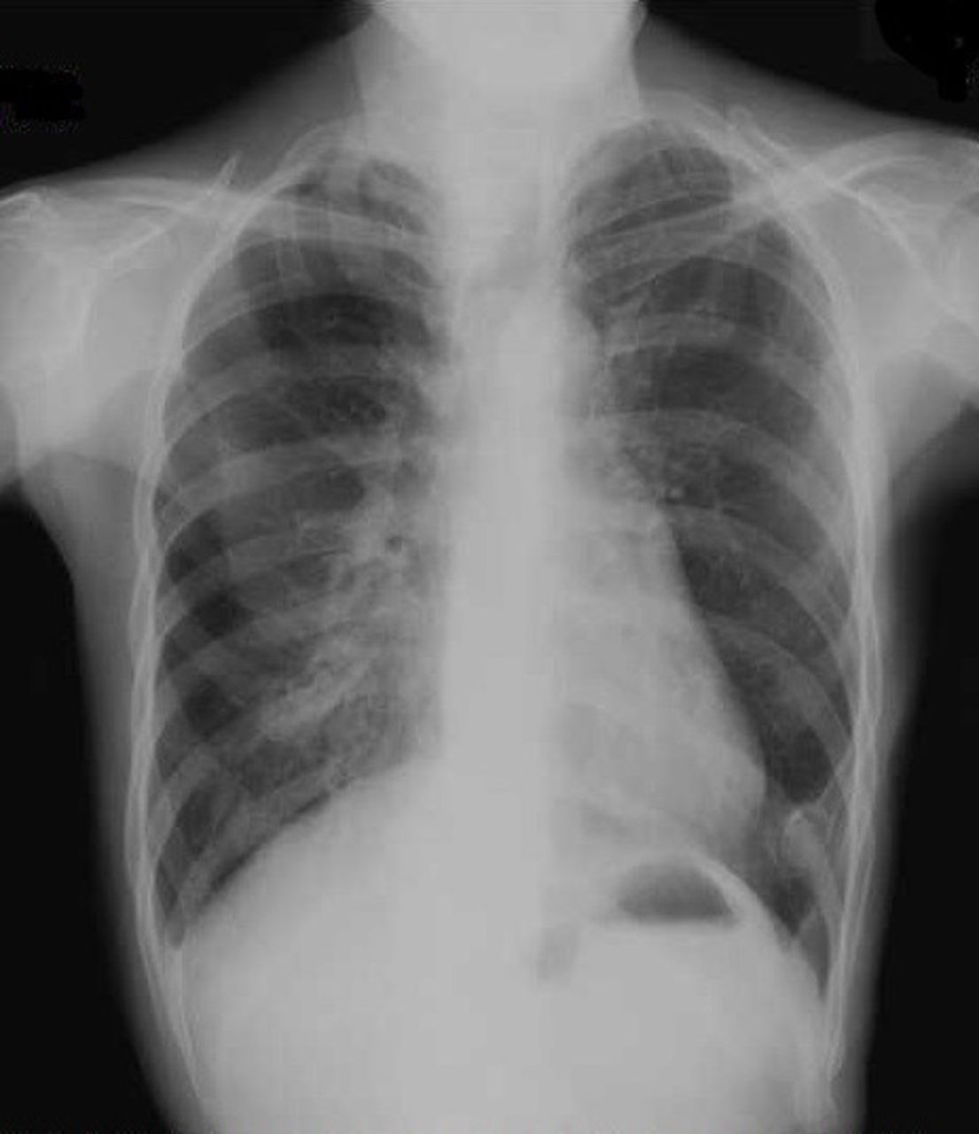
The disrupted narrow trachea and air along the aorta in the mediastinum were observed in chest radiograph.

Sagittal image of CT revealed abscess of 13.2 cm long (black arrowhead) down to the aortic arch surrounding the trachea in mediastinum which compressed the trachea. There was air in anterior and posterior mediastinum and behind the aorta (white arrowhead).
Learning points
Descending necrotising mediastinitis (DNM) is a rare but life-threatening disease, and thus, high index of suspicion for this in patients with severe sore throat is essential.
The complications of DNM include sepsis, great vessel erosions, haemorrhage, cardiac or respiratory problems, as well as abscess in the posterior mediastinum that can cause the airway obstruction, leading respiratory arrest.
Rapid tracheal intubation or tracheostomy for the airway management should be considered when stridor is noted in patients with severe sore throat.
References
Footnotes
Contributors RS cared for the patient. RS, TW and YT wrote the paper.
Competing interests None declared.
Patient consent Obtained.
Provenance and peer review Not commissioned; externally peer reviewed.