Article Text

Summary
Fetus-in-fetu (FIF) is a rare entity in which malformed parasitic twin grows inside the body of its twin. It is most commonly presented with mass in the abdomen. We present a case of a 15-year-old boy who presented with abdominal mass since infancy. Radiological investigations are suggestive of FIF. Intraoperatively, malformed fetus in a sac was found and excised. Postoperatively the patient recovers well and was put on follow-up.
- general surgery
- gastrointestinal surgery
This is an Open Access article distributed in accordance with the Creative Commons Attribution Non Commercial (CC BY-NC 4.0) license, which permits others to distribute, remix, adapt, build upon this work non-commercially, and license their derivative works on different terms, provided the original work is properly cited and the use is non-commercial. See: http://creativecommons.org/licenses/by-nc/4.0/
Statistics from Altmetric.com
Background
Fetus-in-fetu (FIF) is an uncommon pathology with the incidence of 1 per 500 000 births.1 There are many theories proposed with regard to the aetiology of FIF. One of the theories which is increasingly proposed in the recent literature is that the FIF is a diamniotic, monochorionic, monozygotic twin that becomes incorporated into the body of the host twin after anastomosis of the vitelline circulation.2 Other theories include demised multiple pregnancy3 and variant of mature teratoma.4 Most common presentation is a mass in the abdomen.5 Other sites include intracranial, mediastinum, sacrum, scrotum and mouth.6 It can also present with a mass effect. Diagnosis can be made via X-ray, ultrasound, CT scan and MRI. Some cases were diagnosed after prenatal screening.5 Another diagnostic modality (molecular analysis) is using an informative genetic marker for uniparental isodisomy of chromosomes 14 and 15. If there is no genetic difference between the host and the fetiform mass, then it is diagnostic of FIF.5 FIF has to be distinguished from teratoma as the latter has the potential to recur though FIF with malignant recurrence has been reported. Complete excision of the mass is curative and will help in ruling out teratoma with malignant elements.
Case presentation
A 15-year-old boy presented to our hospital with history of abdominal swelling since birth which progressively enlarged recently. He also complaints of abdominal pain and was unable to tolerate orally for 1 week prior to admission. On examination, there was tender, hard mass over central part of abdomen.
Investigations
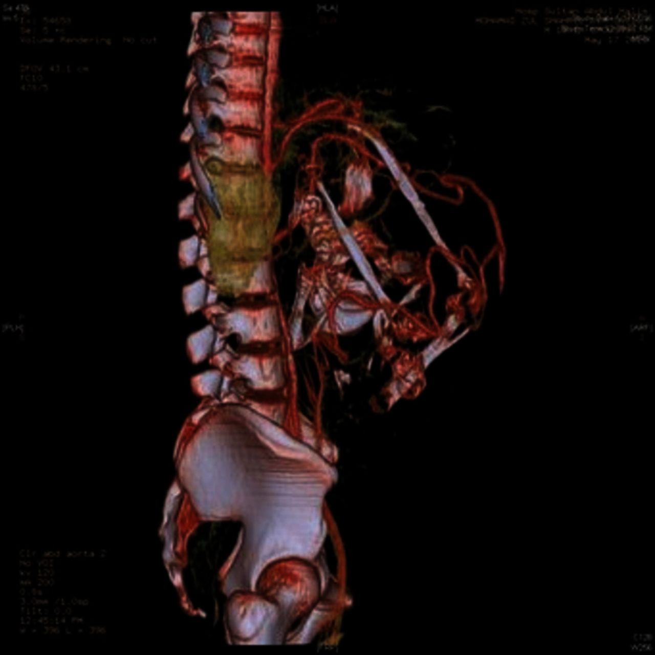
CT abdomen and pelvis revealed a huge intra-abdominal mass 14×18.5×23.8 cm that extends up to the subhepatic region with inferior extension into anterior pelvis. Components in favour of a fetus that were seen within the mass include deformed skull, vertebral body and long bones (figure 1). CT angiography was done to localise the feeding vessels involved. CT angiography shows multiple small arteries supplying the wall of the mass. No main arterial pedicle detected (figure 2). No tumour markers were taken preoperatively.

CT abdomen showing an intra-abdominal mass with components in favour of a fetus within the mass, including deformed skull, vertebra body and long bones. Arrow shows vertebra body.

CT angiography of the abdomen and the pelvis showing multiple small arterial supplying the wall. No main arterial pedicle detected.
Differential diagnosis
The differential diagnosis of this condition would be organised teratoma.
Treatment
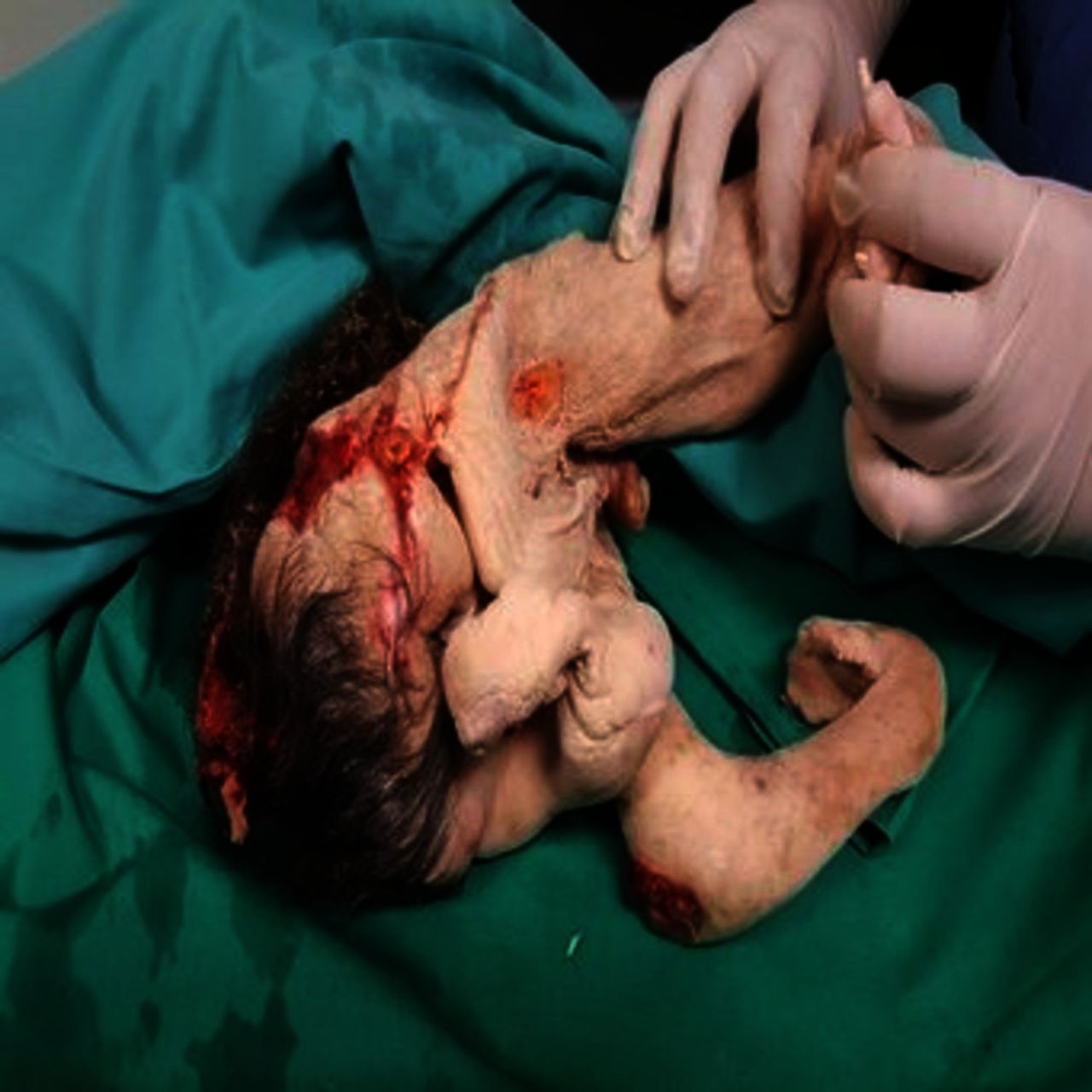
Laparotomy was performed through a midline laparotomy incision with findings of a 15×10 cm intraperitoneal mass with multiple huge feeding vessels densely attached to small bowel mesentery. The mass was perforated over pelvic region with 1000 cc pus aspirated. A gestational sac weighing 2.5 kg was delivered, and when opened, a 1.6 kg non-viable baby noted with shortened and malformed upper and lower limbs, hypoplastic trunk, long hair, developed male genitalia with pubic hair, imperforated anus, fused malformed eyes, vertebra and normal baby skin covered with vernix caecosa. There was no mouth, umbilical cord or placenta (figure 3).

Malformed fetus with hypoplastic trunk, long hair, developed male genitalia, fused eyes and normal baby skin.
Outcome and follow-up
Postoperatively, the baby was returned to the family for ritual funeral as requested by family. Histopathological examination (HPE) of the sac enveloping the fetus revealed a fibrous cyst wall with focal area lining of mature squamous epithelium with many hair shafts and some muscle in subepithelial stroma. The postoperative period of the patient was uneventful. Alpha-feto protein (AFP) taken 3 months postoperation was normal and the patient has no active complaints during subsequent follow ups.
Discussion
It is still a controversy as to whether FIF is an entity on its own or is a highly organised teratoma. Teratoma is a true neoplasm composed of multiple tissues foreign to the part in which it arises.2 Some writers emphasise the presence of vertebral column as distinct feature in diagnosing FIF.5 7 However, there are reports of FIF with differentiation of tissues without vertebral column.8 In our case, although only HPE of the sac was obtained as the baby was returned to family members after the operation for funeral according to local custom, the diagnosis of FIF was confirmed via the presence of vertebral columns in CT abdomen.
Treatment of FIF is surgical. Although benign, complete excision of FIF together with the capsule is crucial as there is a possibility of malignant recurrence if any of the tissue is not completely excised.4 Surgical excision also allows for relieve of obstruction and prevention from further compression.9
The operation to remove FIF is a challenging operation as the mass is highly vascular with multiple feeding vessels. The big size of the FIF has made surgery more difficult as there is high possibility of injuring surrounding structures. CT angiography can help in identifying obvious vascularity and help in assessing the feasibility of doing such cases in the involved centre.10 In our case, CT angiography was done as this is a rare case and the first one encountered in our centre. With limited experience in handling such cases, CT angiography has given a rough idea on the possible difficulties that may be encountered by us intraoperatively especially with regards to vascular supply of the mass.
Learning points
Mass per abdomen with axial skeleton or long bone seen during radiological must raise suspicion of fetus-in-fetu (FIF).
Operation to remove the mass must involve careful planning as there is high possibility of bleeding intraoperatively as the mass is highly vascularised. This involved CT angiography to review the feeding vessel.
FIF must be differentiated from teratoma as there is a risk of recurrence in teratoma.
Further follow up is needed as although the mass is proven to be FIF, there is still a small risk of malignant recurrence especially if there is inadequate removal of sac during operation.
Footnotes
Contributors RY: gave the idea for the article; guarantor. AZM, DZHAJ and NJ: performed literature review. All authors: wrote the article and identified and managed the case.
Competing interests None declared.
Patient consent Guardian consent obtained.
Provenance and peer review Not commissioned; externally peer reviewed.