Article Text

Statistics from Altmetric.com
- Interventional Cardiology
- Clinical Diagnostic Tests
- Diabetes
- Peripheral Nerve Disease
- Physiotherapy (rehabilitation)
Description
A 45-year-old woman with end-stage renal disease and severe diabetes mellitus (haemoglobin A1c (HbA1c) 9.5%) suffered from critical limb ischaemia (CLI). Her right ankle brachial pressure index (ABPI) was 0.77; however, her left ABPI was immeasurable with flat waveform. Additionally, skin perfusion pressure was 34 mm Hg on the left dorsal and 22 mm Hg on the left planter. The angiography showed chronic total occlusion (CTO) of the dorsal pedis artery (DPA) (figure 1, left panel). We electively performed endovascular therapy. The soft wire smoothly passed the CTO of the DPA. We dilated the CTO with a 2.0 mm balloon several times; however, blood flow could not be restored. This discrepancy suggested an unusual CTO. Intravascular ultrasound showed an intact DPA. She also had foot drop due to diabetic neuropathy. Since the entry of CTO was at the dorsal ankle joint, this joint abnormality was considered the cause of the CTO. Manual dorsiflexion of the left ankle joint released the obstruction, ‘pseudo CTO’ (figure 1, right panel). Her left ABPI improved to 1.18 with dorsiflexion. We concluded that the foot drop caused her limb ischaemia.
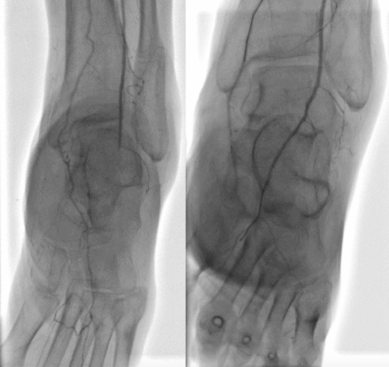
Left panel: the dorsal pedis artery was occluded abruptly like chronic total occlusion at the left ankle joint level. Right panel: manual dorsiflexion relieved the obstruction.
‘External revascularisation’ is one of the CLI treatments. We used total contact cast therapy1 and negative pressure wound therapy to maintain dorsiflexion of her ankle angle and started non-weight-bearing gait training simultaneously (figure 2). Her wound was cured in 2 months. To the best of our knowledge, this is the first report describing the obstruction of the DPA due to foot drop. Foot drop is a common disease whose most frequent cause is a peroneal neuropathy. However, diabetes mellitus is also one of the causes of this disease.2 We need to consider various aetiologies of vascular obstruction and the appropriate therapy for patients with foot drop.

The combination of total contact cast therapy and negative pressure wound therapy. The former keeps the ankle right angle. The latter contributes to early wound healing.
Learning points
Foot drop, a concomitant disorder of diabetes mellitus, causes the occlusion of the dorsal pedis artery. Additionally, ankle dorsiflexion leads to external revascularisation, not internal intervention.
The combination of total contact cast therapy and negative pressure wound therapy is reasonable for keeping dorsiflexion and non-weight-bearing, which contributes to a patient’s cure and early rehabilitation.
The strategy of limb salvage is wide ranging so that many professions must work together as a team.
References
Footnotes
Contributors NK wrote mainly this manuscript. NK, MA, HA and MS designed this concept. HA and MS supervised it.
Competing interests None declared.
Patient consent Obtained.
Provenance and peer review Not commissioned; externally peer reviewed.