Article Text

Statistics from Altmetric.com
Description
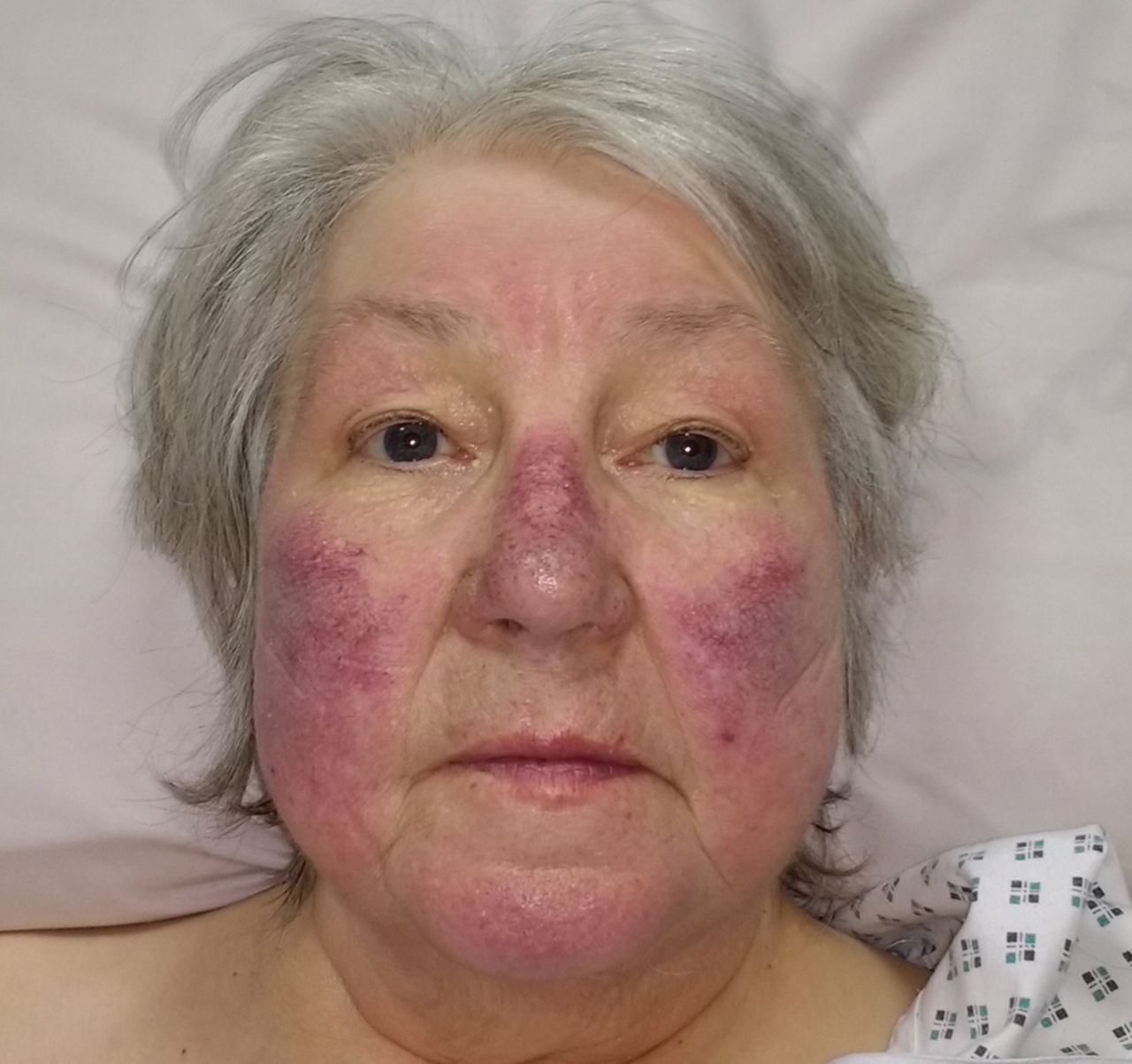
Malar rash is a fixed erythema involving the facial cheeks and nasal bridge with sparing of the nasolabial folds. It has a classic butterfly appearance (figure 1). Its causes include local and systemic diseases, including rosacea, erysipelas and systemic lupus erythematosus (SLE).1 Rarely it occurs due to mitral stenosis with reduced cardiac output and pulmonary hypertension.2 Its association with severe pulmonary hypertension from chronic obstructive pulmonary disease (COPD) is not commonly described.
We recently admitted a 76-year-old woman to our intensive care unit with dyspnoea and cough on a background of COPD. On examination she had a malar rash (figure 1) in association with a respiratory rate of 24 breaths/min, raised jugular venous pressure, peripheral oedema and signs of peripheral venous congestion including leg ulcers and hyperpigmentation of her extremities. Admitting arterial blood gas showed pO2 of 9 kPa on 80% oxygen, pCO2 of 5.5 kPa, pH 7.4 and HCO3 of 27.2 mmol/L. N-terminal pro b-type natriuretic peptide was 18 000 pg/mL. Chest X-ray showed emphysematous lungs and CT pulmonary angiography was negative for pulmonary embolism. Autoantibody screen was negative and physical examination revealed no other features of SLE.

Erythematosus malar rash.
A transthoracic echocardiogram showed severe tricuspid regurgitation with right ventricle pressure approximately 125 mm Hg, dilated right ventricle and impaired left ventricle systolic function with ejection fraction of 35% and no evidence of mitral stenosis. She was managed successfully with a combination of oxygen, diuretics, digoxin, non-invasive ventilation and antibiotics.
Pulmonary hypertension secondary to COPD is common, associated with reduced survival and may worsen during exacerbations. Rarely severe pulmonary hypertension may occur in association with relatively preserved lung function in COPD, and other causes should be considered.3
Learning points
Malar rash can be due to local and systemic disease.
The association of a malar rash with severe pulmonary hypertension should be considered in patients with risk factors, including chronic obstructive pulmonary disease (COPD).
Diagnosis of pulmonary hypertension in COPD has important treatment and prognostic implications.
Footnotes
Contributors RF-O, OT and BB drafted and wrote the manuscript.
Competing interests None declared.
Patient consent Obtained.
Provenance and peer review Not commissioned; externally peer reviewed.