Article Text

Statistics from Altmetric.com
Description
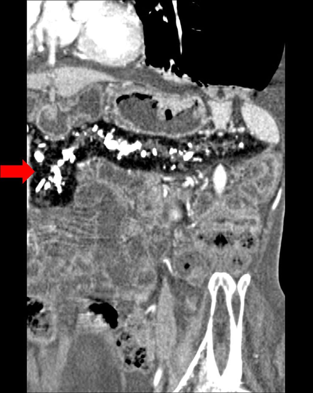
A 31-year-old male patient presented with a history of chronic abdominal pain and progressive loss of weight. Patient also had massive steatorrhea and had been a chronic alcoholic. There was no history of diabetes mellitus, tuberculosis or hypertension. Laboratory investigations revealed profound hypoproteinaemia. Patient underwent a contrast-enhanced CT of the abdomen. It demonstrated a striking ‘dark’ pancreas showing an attenuation of −88 Hounsfield units corresponding to fat (figure 1). No obvious enhancing solid component was seen. Careful review of the multiplanar CT reconstruction images confirmed the presence of dilated pancreatic duct with multiple intraductal calculi (figure 2). The CT findings were diagnostic of total pancreatic lipomatosis secondary to obstructed pancreatic ductal system by calculi/chronic calcific pancreatitis. Patient was managed conservatively using pancreatic enzyme replacement therapy.

Axial contrast-enhanced CT image shows the dark, hypoattenuating pancreatic parenchyma corresponding to fat. Note the intraductal calculi (arrow) obstructing the main pancreatic duct. Bilateral pleural effusion and ascites are also seen secondary to hypoproteinaemia.

Curved multiplanar reconstructed image shows entire pancreas replaced by fat suggestive of total pancreatic lipomatosis (arrow).
Pancreatic lipomatosis refers to replacement of exocrine pancreas with fat. It is synonymous to lipomatous pseudohypertrophy or pancreatic steatosis. It may range from focal to diffuse accumulation of fat in the pancreas. Focal replacement does not carry much clinical significance; however, total involvement affects the pancreatic exocrine function resulting in malabsorption.1 It is seen in diseases like diabetes mellitus, hereditary and chronic pancreatitis, pancreatic duct obstruction by calculus or tumour, Cushing's disease, steroid therapy, haemochromatosis, viral infections and an increasing incidence in patients with obesity and metabolic syndrome.2 It can also be seen in association with few congenital diseases, for instance, cystic fibrosis, Shwachman-Diamond syndrome and Pearson syndrome (mitochondrial). Pancreatic lipomatosis is easily demonstrable on common imaging techniques such as CT and MRI, which are diagnostic for the entity. In addition, CT can provide a clue in detecting specific aetiology causing lipomatosis, for example, an obstructive intraductal calculus or tumour. Though MRI is the best imaging technique to demonstrate the entity, present generation CT scanners providing high-resolution images can demonstrate the condition with equal sensitivity and specificity. It is important to confirm that the pancreatic duct is seen on CT/MRI images, which helps in distinguishing this entity from pancreatic agenesis.3 With its typical imaging features verifiable, CT can reliably be used as imaging modality of choice to confidently diagnose or exclude the pancreatic lipomatosis. Pancreatic enzyme replacement therapy in combination with dietary counselling is the mainstay of the management in these patients.
Learning points
Total pancreatic lipomatosis affects the exocrine pancreatic function and leads to malabsorption.
CT can reliably diagnose or exclude the pancreatic lipomatosis and may suggest a possible specific aetiology.
It is important to confirm that the pancreatic duct is seen, which helps in distinguishing the entity from pancreatic agenesis.
Footnotes
Contributors PN and SM have been equally involved in actively performing the CT of the index case and in planning, conducting and reporting of the work described in the article.
Competing interests None declared.
Patient consent Obtained.
Provenance and peer review Not commissioned; externally peer reviewed.