Article Text

Statistics from Altmetric.com
Description
A Caucasian woman aged 56 years presented to emergency room (ER) department with leucorrhoea and fever since 48 hours. She was a smoker, had no diabetes history, no prosthetic material and denied use of injected drugs.
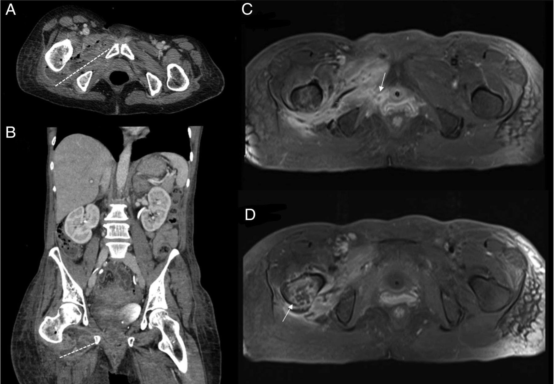
A month before, the patient had a closed inguinal trauma due to fall from height with a muscle strain of the anterior right thigh. Despite rest and analgesics, she went to ER several times because of progressive local pain and swelling. After 3 weeks, a local ultrasound scan showed a 3 cm size haematoma associated with probable rupture of obturator internus and rectus femoris muscles. On the following days, she developed fetid leucorrhoea associated with movements and compression of the anterior thigh. On physical examination and analyses, she had sepsis criteria and the abdomen/pelvic CT scan (figure1A, B) and MRI (figure1C, D) showed an abscess of 105×25mm size, complicated with osteomyelitis of the femur and fistulisation to the vagina through obturador foramen. The patient was treated with empiric large-spectrum antibiotics (piperacillin/tazobactam and linezolid) and surgical drainage with soft-tissue debridement was performed. Anaerobic agents and a Streptococcus specimen were found on the exudate material. Blood cultures and HIV test were negative. One month has passed and the patient is clinically and radiologically improving. The antimicrobial therapy will be maintained according to her medical course.
Abdominal and pelvic CT scan, axial (A) and coronal (B) views, showing a multilobulated heterogeneous abscess of the right thigh containing gas, with 105×25×52 mm size (intermittent white line). (C and D) Magnetic resonance axial T1-weighted postgadolinium images revealing the abscess of the right thigh with fistulae to vagina (C, white arrow) and signs of osteomyelitis of the right greater trochanter and femoral head (D, white arrow).
In this patient, the gradual symptoms and the closed trauma misled the physicians, leading to delay of the diagnosis. The acute osteomyelitis was secondary to a contiguous spread of the infected haematoma and, as expected in these cases, polymicrobial population was identified.1 The extension of the infection with fistulisation is an unusual feature in a patient with no immunosuppressive condition.
Learning points
Delay in the diagnosis of soft tissue infection can lead to catastrophic consequences, such as contiguous spread of infection to bone and fistulisation, even in immunocompetent patients.
Since the key of successful management of osteomyelitis is early diagnosis, physicians must keep high suspicion in patients with persistent pain after trauma.
Acute osteomyelitis may respond to antibiotics alone and the duration of treatment should be guided by clinical evolution.
Reference
Footnotes
Twitter Follow Carolina Ourique @Carolina Ourique
Contributors CO and FR-C made the diagnosis. All authors were involved in management of the patient, wrote the manuscript and performed the literature research. PTR corrected the manuscript and gave conceptual advice. All authors read and approved the final version of the manuscript.
Competing interests None declared.
Patient consent Obtained.
Provenance and peer review Not commissioned; externally peer reviewed.