Article Text

Statistics from Altmetric.com
Description
A 58-year-old man presented with history of irritative lower urinary tract symptoms (daytime frequency and nocturia) for 3 months. Digital rectal examination revealed grade 2 prostatomegaly and consistency was hard. Serum prostate-specific antigen (PSA) was 2.7 ng/mL. Serum creatinine was 1.2 mg/dL. Transrectal ultrasonography (TRUS) revealed a prostate size of 54×47×56 mm with hypoechoic nodular appearance at superior part of the left lobe. Ultrasound scan of the abdomen was normal. Urinary microscopy revealed 1–2 pus cells and 3–4 erythrocytes. TRUS-guided 12-core biopsy was performed. Microscopic examination of all the biopsy cores revealed fused glandular structures with focal cribriform pattern (Gleason pattern 4+4=8), involving >80% of the prostatic cores (figures 1 and 2). Perineural invasion was evident. Individual cells were polygonal with voluminous foamy cytoplasm, low nucleo-cytoplasmic ratio, pyknotic nuclei and inconspicuous nucleoli. Tumour cells were immunoreactive for PSA while p63 and 34βE12 confirmed the absence of basal cell layer (figure 3). Radical prostatectomy was performed. PSA nadir (0.01 ng/mL) was achieved at 1 month post procedure. The patient is doing well after 12 months of follow-up with no evidence of recurrent or residual disease.

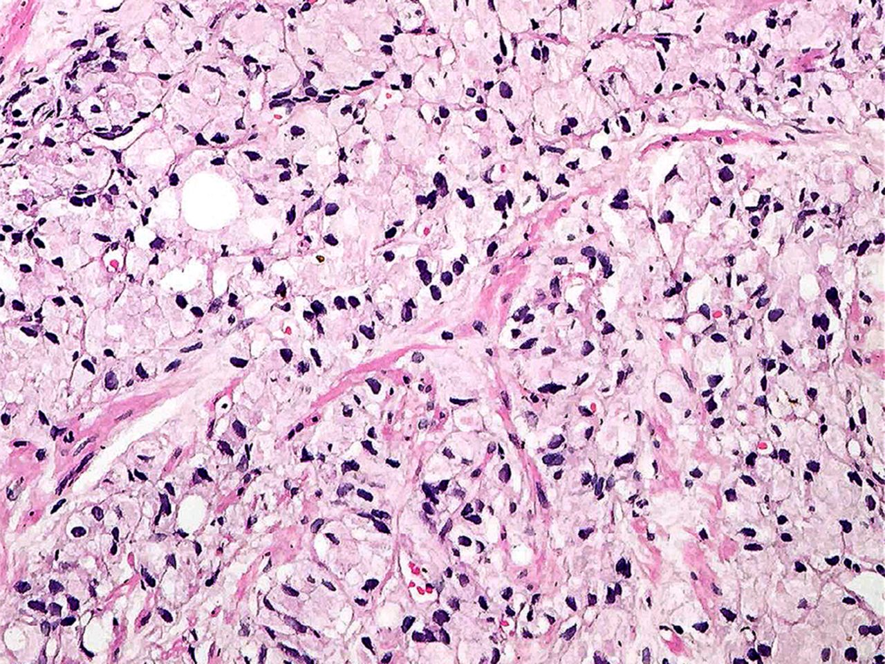
Needle core biopsy showing diffuse infiltration by a tumour composed of crowded glands lined by cells showing voluminous foamy cytoplasm. The overall Gleason score was 4+4=8.

Foamy gland carcinoma showing fused glandular structures with cells having abundant foamy cytoplasm, small pyknotic bland looking nuclei and absent nucleoli.

Immunohistochemical stains. (A) Tumour cells staining positive with PSA. (B) p63 demonstrates the absence of basal cell layer in the tumour. (C) 34βE12 shows the absence of basal cell layer in the tumour.
Foamy gland variant of prostatic adenocarcinoma was first described in 1996 by Nelson and Epstein.1 It occurs in a significant proportion (17%) of cases of carcinoma prostate diagnosed on needle biopsy.2 Age at presentation ranges from 50 to 78 years. The tumour cells are deceptively benign and characteristically have abundant foamy cytoplasm, bland pyknotic nuclei and inconspicuous nucleoli. International Society of Urologic Pathology (ISUP) recommends grading of these carcinomas based on the underlying architectural pattern.3 Prognosis depends on the Gleason score and the presence or absence of perineural or extra prostatic extension. Recognition of this variant poses a challenge for an uropathologist and can be missed on needle biopsy.
Learning points
Foamy gland variant of prostatic adenocarcinoma mimics benign glands.
It is important for the pathologist to be aware of this entity as foamy gland carcinoma in pure form can be easily missed on needle biopsy.
Gleason scoring of this variant is performed by the underlying architectural pattern.
Footnotes
Contributors AA involved in planning, conduct, reporting, conception and design, acquisition of data or analysis. RJ is responsible for conduct, reporting, conception and design, interpretation of data. NA reviewed the literature and involved in writing the manuscript. NH is responsible for supervision, review of the literature and analysis of data.
Competing interests None declared.
Patient consent Obtained.
Provenance and peer review Not commissioned; externally peer reviewed.