Article Text

Statistics from Altmetric.com
Description
A 19-year-old woman born to non-consanguineous parents presented with primary amenorrhoea. Examination revealed a 1.3 m tall patient with disproportionate dwarfism (arm-span/height—ratio: 0.78); she was polydactyl with hypoplastic/dystrophic nails (figure 1). She had partial anodontia, and abnormally shaded and mal-occluded teeth (figure 2). The rest of the systemic examination was unremarkable. Radiography of the hands revealed fusion of the carpal bones (figure 3). Echocardiography revealed patent foramen ovale with no flow. No haematological/biochemical abnormalities were detected. The patient's history revealed no similar anomaly in the family. The patient could not afford genetic mutation analysis. Her clinical phenotype was characteristic of Ellis-van Creveld (EVC) syndrome. Anatomic evaluation of her reproductive tract, serum prolactin/follicle stimulating hormone/luteinizing hormone/growth hormone assays and brain MRI were normal. The primary amenorrhoea was unexplained despite follow-up for 5 years.

Clinical photograph showing (A) patient height depicted on scale; (B) bilateral polydactyly; (C) hypoplastic/dystrophic nails noted over bilateral thumbs.
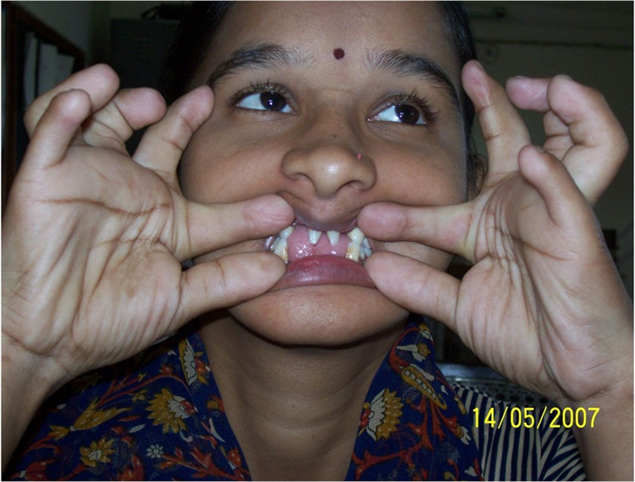
Clinical photograph showing oral abnormalities including partial anodontia in upper and lower jaw, abnormally shaded (yellow stained) teeth and conical incisors showing hand anomalies.

Radiography showing (A) bilateral polydactyly and fusion of carpal bones, namely, capitate and hamate on X-ray of both hands; and (B) partial anodontia and malocclusion of teeth on X-ray the skull.
EVC syndrome is an autosomal recessive disorder with prevalence of 1 in 60 000–150 0001 secondary to mutation in EVC and EVC-2 genes on chromosome 4p16.2 Four distinctive features include (1) chondrodystrophy with average adult height between 109 and 155 cm; (2) polydactyly mostly involving the hands and, rarely, (10%) the feet; (3) ectodermal dysplasia presenting as tooth and nail abnormalities, reported in 93% of published cases. Tooth abnormalities include neonatal/maloccluded/small/abnormally shaded teeth with delayed eruption and partial anodontia. Nail abnormalities include hypoplastic/dysplastic/friable/absent nails; and (4) cardiac abnormalities, seen in 50–60% patients, with atria being commonly involved.2 ,3 Our patient had all four primary manifestations, strongly suggesting the diagnosis despite the absence of genetic testing. Amenorrhoea is rarely reported in EVC syndrome and mostly due to vaginal agenesis.
Learning points
Ellis-van Creveld syndrome is an ectodermal dysplasia.
Cardinal manifestations are chondrodystrophy, polydactyly, tooth or nail abnormalities and atrial defects.
Footnotes
Contributors All the authors were actively involved in managing the patient over the past 8 years. UY drafted the manuscript.
Competing interests None declared.
Patient consent Obtained.
Provenance and peer review Not commissioned; externally peer reviewed.