Article Text

Summary
We describe a rare case of a young woman with a large cystic adenomyotic lesion that was treated laparoscopically. The patient presented with severe dysmenorrhoea refractory to common analgaesics. She was initially diagnosed with right-sided ovarian endometrioma. MRI revealed a cystic lesion of 4 cm attached to the right uterine wall. Under laparoscopic vision, the uterine lesion was identified on the right portion of the uterine fundus close to the round ligament. Monopolar diathermy was used to dissect the lesion. When the incision reached the cystic cavity, dark-brown content flowed from the cyst. After resection was complete, the surgical wound was closed with two-layer interrupted sutures. The patient made a good recovery and was discharged the following day. Since patients with cystic adenomyosis are young, a minimally invasive procedure such as laparoscopic excision is considered optimal. The exact topography of the lesion is crucial in determining the site of the incision.
Statistics from Altmetric.com
Background
We report a rare case of a young woman with a large cystic adenomyotic lesion that was treated laparoscopically. The patient presented with severe dysmenorrhoea and pelvic pain, which was unsuccessfully treated with common analgaesics. Owing to the rarity of such lesions and their close proximity to the ovaries, they are often considered to be endometriotic cysts. Although adenomyosis is often asymptomatic, cystic adenomyosis is generally associated with dysmenorrhoea and/or chronic pelvic pain. Evidence from several case reports suggests that surgical removal of the cyst is the best treatment option to relieve the patient's symptoms. The feasibility of laparoscopic excision of the lesion along with uterine reconstruction is presented in this case. Conservative uterine-sparing surgery for adenomyosis is considered a viable and efficacious method of treatment in young patients who wish to preserve fertility. The successful outcome of this patient adds to what has already been described by others.
Case presentation
A 28-year-old nulligravida woman presented with a history of dysmenorrhoea and pelvic pain for the past year. The patient underwent menarche at the age of 14 and was asymptomatic until the age of 20. Thereafter she was experiencing progressively worsening dysmenorrhoea, which was relieved with mild analgaesics. During the past 12 months, the patient reported gradual increase of the severity and duration of the dysmenorrhoea, lasting until day 7 and 8 of the menstrual cycle. The response to high doses of non-steroidal anti-inflammatory drugs was poor and necessitated hospitalisation on several occasions for intravenous administration of analgesics. The patient came to the emergency unit of the Obstetrics and Gynaecology Department of the University Hospital of Larissa, with severe dysmenorrhoea and lower abdominal pain. She had no sexual history and opted for a transabdominal scan, which revealed a thick-walled hypoechoic mass of 4×3.5 cm in close proximity to the uterus. The mass resembled a right ovarian endometriotic cyst and that was the initial diagnosis. She consented for a transrectal scan, which showed both adnexae and an accessory uterine mass with a haemorrhagic cystic lesion of 4 cm attached to the right uterine wall.
Investigations
Pelvis MRI was then scheduled on a 3T magnet (GE HdX, Milwaukee, USA). Axial, sagittal and coronal T1 and T2-weighted images were obtained before and after intravenous contrast administration of gadobenate dimeglumine (0.1 mmol/kg). A haemorrhagic, well-defined mass was identified on the right side of the uterine wall. The lesion seemed to be an exophytic myometrial mass, 3.7×2.4×3.2 cm in size, presenting with low signal intensity on T2-weighted images and increased signal intensity on T1-weighted images. No contrast enhancement was identified (figure 1A–D). The uterus was arcuate-shaped and adenomyosis of the myometrium was identified (figure 2A, B). The ovaries were clearly identified, along with with several follicles (figure 2C).
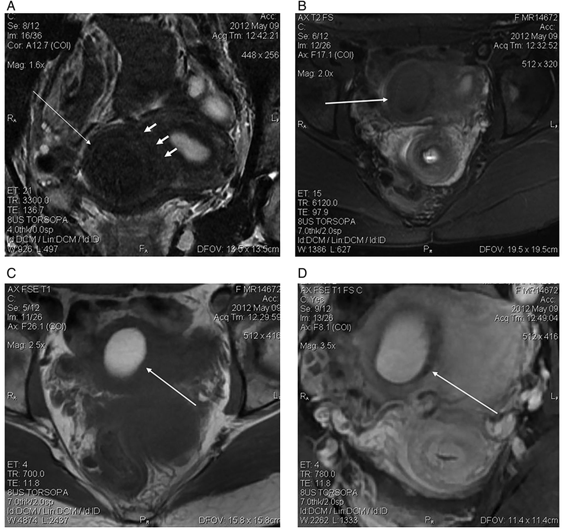
Coronal (A) and axial (B) T2-weighted images showing an exophytic low signal intensity mass originating from the right side of the myometrium. (C) Axial T1-weighted images show increased signal intensity of the lesion, (D) no contrast enhancement is identified. There is no border identified between the lesion and the uterine wall (small arrows).

Sagittal (A) and coronal (B) T2-weighted images of the uterus show adenomyosis of the myometrium and the arcuate shape of the uterus. Coronal T2 images show the ovaries (C) filled with multiple follicles.
Differential diagnosis
Differential diagnosis should include congenital uterine anomalies and endometriotic cysts, which in many cases are in close proximity to the uterine wall.
Treatment
Based on the imaging studies and patient's history, suspicions of cystic adenomyosis were raised. The patient was then advised to undergo laparoscopic excision of the mass. By the time of the operation the patient was sexually active, and a uterine manipulator was used with her consent. The laparoscope was inserted into the abdominal cavity from an 11 mm supra-umbilical incision using an open access technique.1 Three additional trocars were introduced under direct vision. After visual inspection of the pelvic cavity, the site of the uterine incision was determined by visual palpation of the uterus and based on previous sonographic evaluation. Diluted vasopressin was injected (1 mL of 20 U of vasopressin diluted with 40 mL of normal saline) in the myometrium around the affected area. The uterine wall was incised with monopolar current, set at 30 W. The uterine lesion was identified on the right portion of the uterine fundus close to the round ligament (figure 3A, B). When the incision reached the cystic cavity, dark-brown content flowed from the cyst (figure 3C). There was no direct communication between the endometrial cavity and the cystic mass (figure 3D). Similarly to cases with focal or diffuse adenomyosis, the cleavage plane was difficult to identify. Once resection was complete, the surgical wound was closed with deep two-layer interrupted, 1-monocryl sutures. The serosal layer was closed with interrupted monocryl 0 sutures (figure 3F). Care was taken to avoid injury to the right fallopian tube. The specimen was morcellated out of the abdominal cavity. The histopathology of the specimen demonstrated endometrial epithelium and stroma lining the cystic cavity and adenomyosis in the adjacent myometrium confirming the diagnosis of cystic adenomyosis.

(A) Laparoscopic view of the uterine cystic lesion (marked with arrows) located in the right portion of the uterine fundus. Superficial endometriotic lesions are also visible at the uterosacral ligaments. (B) Monopolar diathermy was used to dissect the lesion. (C) Dark-brown content flowed from the cyst when the incision reached the cystic cavity. (D, E) The cystic nodule was removed and had no direct communication with the endometrial cavity. (F) The surgical wound was closed with deep interrupted 1-monocryl sutures in two layers. The serosal layer was closed with interrupted monocryl 0 sutures.
Outcome and follow-up
The patient made a good recovery and was discharged the following day. She was relieved from symptoms and remained asymptomatic 1 year after the surgery.
Discussion
The most common presentation of adenomyosis is in its diffuse form, but it can also present as a focal lesion, which is also defined as adenomyoma. Dilated cystic glands or haemorrhagic foci can be present in the ectopic endometrium resulting in the appearance of myometrial cysts <5 mm, while in a very rare form of adenomyosis, the extensive haemorrhage within the ectopic glands results in the appearance of a lesion characterised as adenomyotic cyst, cystic adenomyosis or cystic adenomyoma.
Cystic adenomyosis represents a rare entity, and is more commonly encountered in younger patients. Based on the patient's age at symptom development, cystic adenomyosis is traditionally classified into two categories: juvenile cystic adenomyosis and adult cystic adenomyosis. For a case to be classified as juvenile cystic adenomyosis, the patient's symptoms should start within 5 years after menarche or ≤18 years of age.2 Adult cystic adenomyoma is usually found in older patients and most often in those over the age of 30. Acién et al3 recently proposed the term accessory and cavitated uterine masses (ACUM) with a functional endometrium these include cases of juvenile and cystic adenomyosis. The inclusion criteria for a uterine cystic lesion to fit in the classification are the following, they should: (1) be an isolated accessory cavitated mass; (2) be in a normal uterus (endometrial lumen), with normal Fallopian tubes and ovaries; (3) be a surgical case with an excised mass and a pathological examination; (4) be an accessory cavity lined by endometrial epithelium with glands and stroma; (5) have a chocolate-brown-coloured fluid content; and (6) have no adenomyosis (if the uterus has been removed), although there could be small foci of adenomyosis in the myometrium adjacent to the accessory cavity.3 In our case, the patient fulfilled all the above criteria, since the histopathological report showed that the lining of the accessory cavity consisted of endometrial epithelium and stroma, however, since this classification is not widely accepted, we considered our case to be a large cystic adenomyotic lesion.
Diagnosis and treatment of these cases pose great difficulties that will be hard to overcome until well-designed studies are launched to guide management.4 It seems that, although adenomyosis is asymptomatic in most cases and represents a histopathological finding in women undergoing hysterectomy, cystic adenomyosis is invariably associated with pelvic pain and dysmenorrhoea.5 Transvaginal sonography is usually the first choice of image modality when investigating cases of pelvic pain, but accurate diagnosis of cystic adenomyosis is not always possible, since correct recognition of the specific sonographic features of the disease is difficult. Sonographic localisation of the myometrium and the possible relation of the lesion to the endometrial cavity can be challenging. In several cases, lesions are assumed to be adnexal. Owing to its hyperechoic content, the cystic lesion in our patient was initially considered to be an endometriotic cyst of the right ovary. The exact origin of the mass was specified by means of transrectal scan and MRI. MRI is most helpful in these cases, since it is less observer dependent and thus considered a more accurate non-invasive technique for the diagnosis of adenomyosis.6 Three-dimensional sonography has been proposed as an accurate diagnostic aid in determining the myometrial borders of adenomyosis preoperatively, however, such a modality was not performed in our patient.
Adenomyosis is a disease that grows into the myometrial layer of the uterus, affecting its structure and texture. Conversely to laparoscopic removal of myomas, where the cleavage plane is clear, excision of an adenomyotic lesion is difficult. Primary consideration in these cases is the removal of the whole of the adenomyosis, if possible, while preserving as much unaffected myometrium as possible. Since many patients with cystic adenomyosis are young, a minimally invasive procedure such as laparoscopic excision is considered preferable. However, many cases have been treated by laparotomy mainly due the difficulty in determining the exact location of the mass and the high expertise needed to safely remove the entire lesion by laparoscopy. The main criterion on identifying the defective tissue during laparoscopy is the macroscopic appearance of the lesion. Adenomyotic tissue is paler, less vascular and bleeds less due to fibrosis, contrary to unaffected tissue, which is redder and more haemorrhagic. No dead space should be left during suturing and unnecessary use of diathermy should be avoided. In cases where the endometrial cavity has been entered, this should be closed separately. Two layers of interrupted sutures were employed in our case, which assured good approximation of the uterine surfaces. Since preserving fertility is the ultimate goal of this surgery, meticulous reconstruction of the uterus should be performed in order to minimise the risk of uterine rupture in a subsequent pregnancy.
Removal of the specimen from the abdominal cavity is another issue that has recently raised concerns regarding the spread of tissue with malignant potential during morcellation. Although our specimen was morcellated, and the surgery took place a while ago, in view of the recent Food and Drug Administration (FDA) comments, we also feel that the use of an endobag presents the safer choice in these cases.
Partial resection of adenomyosis during laparoscopy or laparotomy is an accepted mode of treatment in cases of cystic adenomyosis.7 The size and location of the lesion and the expertise of the surgeon are the two major determinants of the surgical route. Safety issues that arise by the wide use of this conservative uterine-sparing surgery, should be addressed promptly by large-scale well-designed studies.
Patient's perspective
I used to think that I would be in pain all my life. My doctors said that it was normal to feel a degree of pain during my period and that I had to remove the endometriotic cyst at some point. They said that removal of the cyst would not relieve my symptoms. Since the suspicion of cystic adenomyosis was raised and a possible treatment was offered I was keen on having it done despite the possible risk. I am a different person now. My periods are without pain. Occasionally, I notice a slight spotting at the end of my period, but my doctor assured me that everything is well.
Learning points
Αlthough adenomyosis is, in most cases, asymptomatic and represents a histopathological finding in women undergoing hysterectomy, cystic adenomyosis is invariably associated with pelvic pain and dysmenorrhoea.
Diagnosis of cystic adenomyosis is not always possible. Sonographic localisation of the myometrium and the possible relation of the lesion to the endometrial cavity can be challenging.
Adenomyosis is a disease that grows into the myometrial layer of the uterus, affecting its structure and texture. Conversely to laparoscopic removal of myomas, where the cleavage plane is clear, excision of an adenomyotic lesion is difficult.
Since many patients with cystic adenomyosis are young, a minimally invasive procedure, such as laparoscopic excision, is considered preferable.
Footnotes
Contributors OK was responsible for diagnosis of the patient and writing of the manuscript. EK was responsible for diagnosis of the patient. AD was responsible for writing and editing of the manuscript. GP was the surgeon who carried out the procedure.
Competing interests None declared.
Patient consent Obtained.
Provenance and peer review Not commissioned; externally peer reviewed.