Article Text

Statistics from Altmetric.com
Description
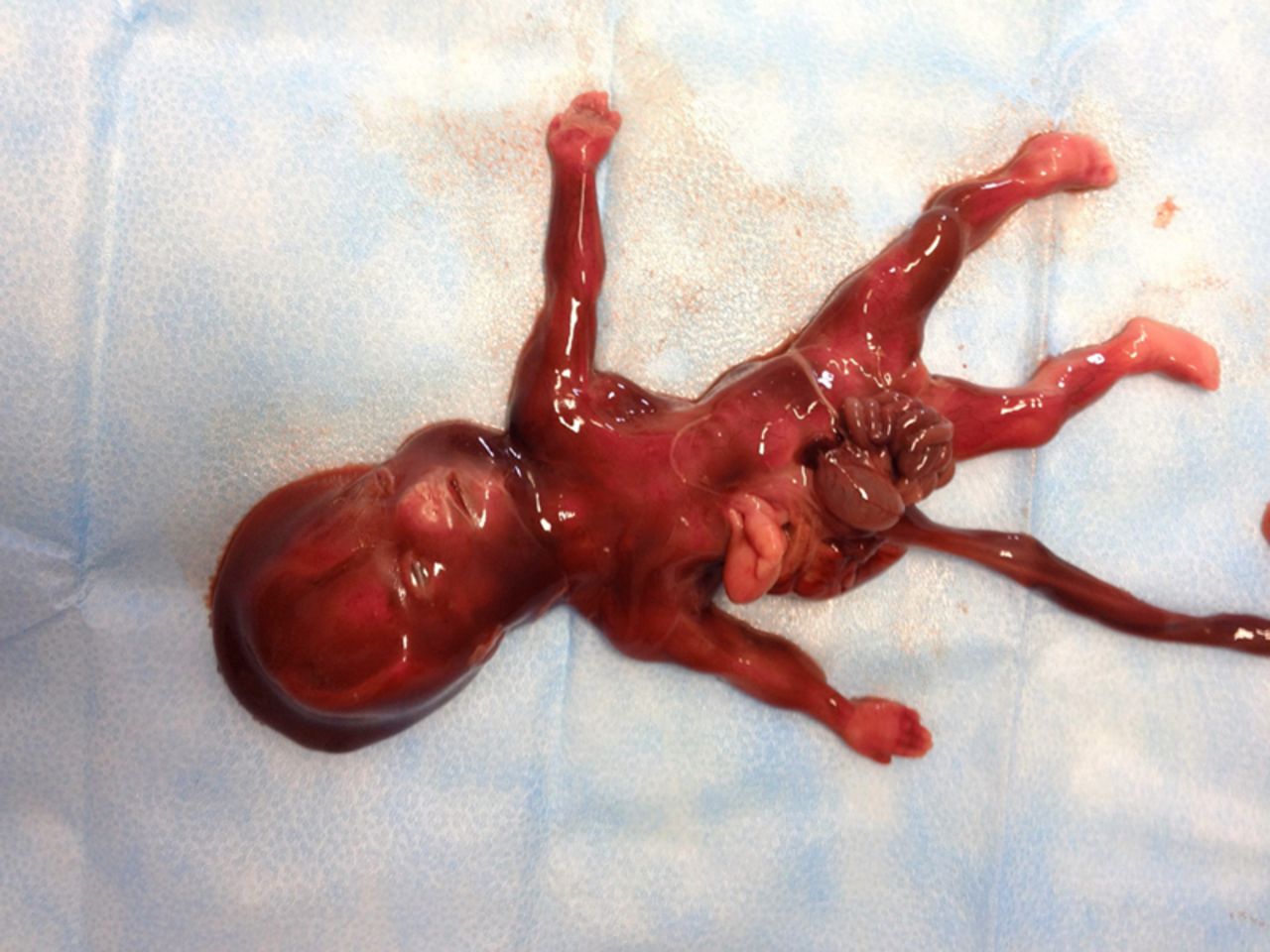
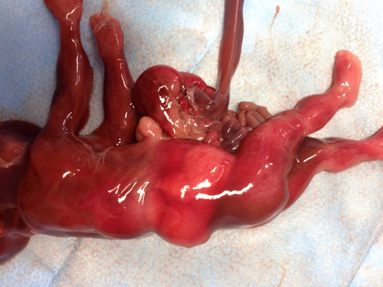
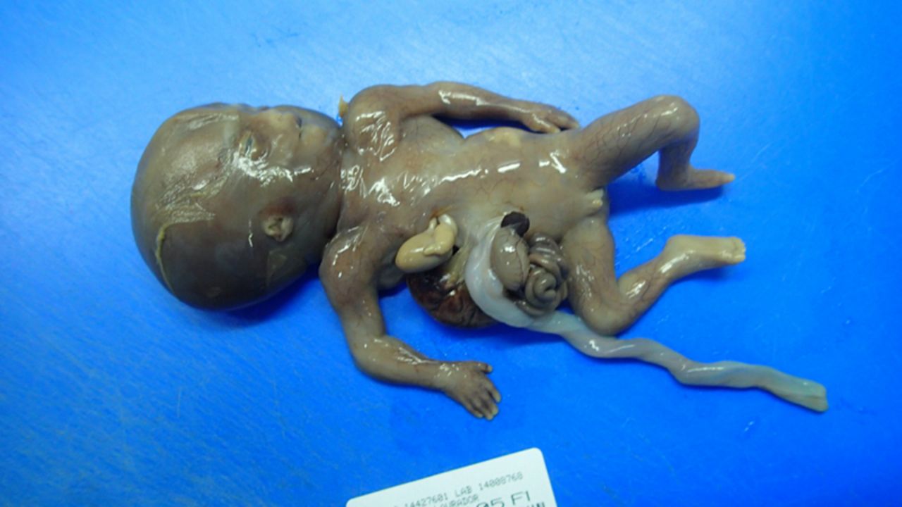
A healthy 25-year-old primigravida, without a history of teratogenic exposition, presented to our hospital with a first trimester routine ultrasonographic examination showing two major fetal defects: gastrosquisis (figure 1 and video 1) and ectopia cordis (video 2). At 16 weeks of gestation a detailed ultrasound confirmed the suspected abnormalities together with sternal and diaphragm defects, and pentalogy of Cantrell was suspected. An amniocentesis and termination of the pregnancy were considered and accepted by the couple. After expulsion, we identified a dead fetus (figure 2) weighing 113 g, with a thoracoabdominal wall defect with ectopia cordis and evisceration of stomach, liver, intestines and spleen (figures 3 and 4). The anatomopathological examination (figures 5 and 6) recognised additional cardiac defects, which provided the final diagnosis of pentalogy of Cantrell: absent pericardium and diaphragm, lower sternal defect, transposition of great vessels, severe right ventricle hypoplasia, enlarged left cavities and intraventricular communication. No other malformations or chromosomal abnormalities were documented. First described in 1958 by Cantrell, pentalogy of Cantrell (OMIM 313850) is an extremely rare and usually lethal congenital malformation, with an estimated incidence from 5.5 to 7.9 per million live births. The full spectrum is rarely reported and consists of five anomalies: anterior abdominal wall defect, anterior diaphragmatic hernia, sternal cleft, ectopia cordis and intracardiac abnormalities.1 A possible pathogenesis involves a defect in the differentiation of the intraembryonic mesoderm between 14 and 18 days after conception, so a first trimester ultrasonography is indispensable for the prenatal diagnosis.2 Prognosis is poor, depending on the extent of the defects. Few live births of the complete pentalogy have been described.3
Learning points
The hallmark of this syndrome is ectopia cordis with anterior abdominal wall defect. In the presence of these abnormalities, obstetricians should consider pentalogy of Cantrell.
Cantrell’s syndrome can be diagnosed as early as the first trimester using a two-dimensional (2D) ultrasound. A 3D ultrasound can help to enhance the visualisation of the fetal anomalies.
Pentalogy of Cantrell must be adequately evaluated for appropriate prenatal counselling and postnatal management of individual cases. Survival depends on the number and severity of the defects so an early diagnosis gives the parents an option of termination of pregnancy.

Ultrasound at 14 weeks revealing gastrosquisis.

Delivered dead fetus.

Thoracic defect with ectopia cordis plus abdominal wall defect with severe gastrosquisis: liver, stomach, intestines and spleen.

Ectopia cordis and severe gastrosquisis.

Anatomopathological examination of the fetus.

Anatomopathological examination of the fetus.
Footnotes
Competing interests None.
Patient consent None.
Provenance and peer review Not commissioned; externally peer reviewed.