Article Text

Statistics from Altmetric.com
Description
A 71-year-old woman was referred due to chest discomfort. She had normal breath sounds with normal pulmonary functions. A chest roentgenogram was also normal, while a transverse thin section CT image (1 mm section thickness) showed bronchial wall thickening with calcifications in the left lower bronchus (arrows, figure 1). A reconstructed coronal CT image in the lung window showed focal narrowing of the left lower bronchus (arrows, figure 2). Bronchoscopy revealed diffuse redness and thickening of the bronchial mucosa with mild stenosis of the left lower bronchus (figure 3). The pathological diagnosis of a transbronchial biopsy was neurinoma composed of spindle cells with a palisading pattern (figure 4) containing S-100 protein-positive cells (inset), which was distinguishable from leiomyoma or meningioma by the absence of abundant elongated eosinophilic cytoplasm or inflammatory infiltration of plasma cells. The patient had neither acoustic neurinoma nor cafe-au-lait spots on the skin. She is considered to be at risk of bronchial stenosis in the future, and it may be necessary to consider bronchoscopic argon plasma coagulation1 or left lower sleeve lobectomy. However, the CT findings have remained stable for 10 years, and watchful observation has proven to be a valid choice in this case. Primary endobronchial neurinomas are extremely uncommon, constituting approximately 2% of benign tracheobronchial tumours.2 Surgery had been chosen in most reported cases of endobronchial neurinoma1 ,3; however, an awareness of the possibility of this disease and proper management based on each patient's clinical condition is required. We presented a rare case of primary endobronchial neurinoma with fascinating imaging findings.3
Learning points
-
Primary endobronchial neurinoma is one of uncommon benign tracheobronchial tumours.
-
A reconstructed coronal CT image in the lung window is impressive for this disease.
-
The pathological diagnosis is necessary for distinguishing from other tumours, such as leiomyoma or meningioma.
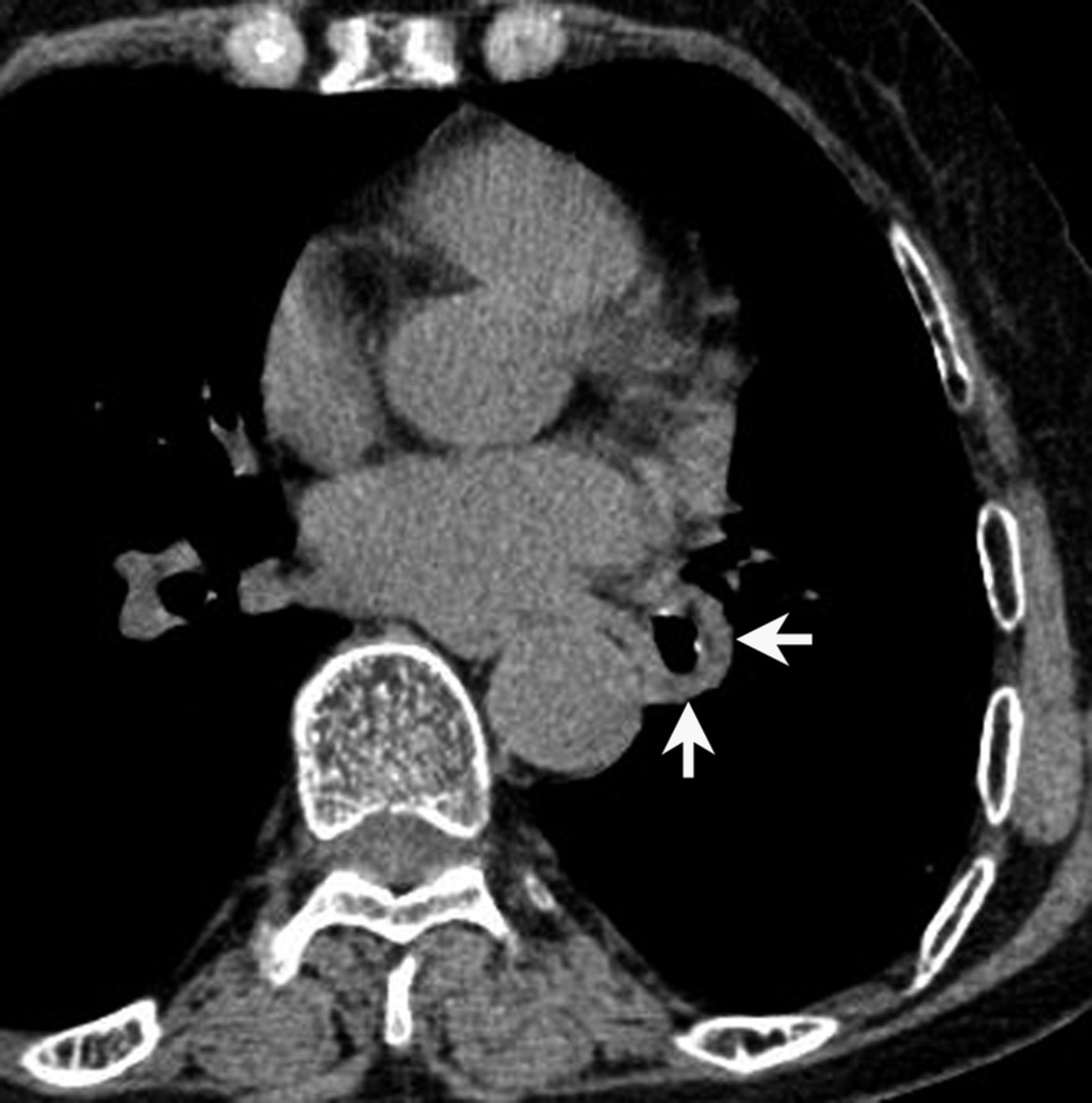
A transverse thin section CT image (1 mm section thickness) showing bronchial wall thickening of the left lower bronchus (arrows).

A reconstructed coronal CT image in the lung window showing the focal narrowing of the left lower bronchus (arrows).
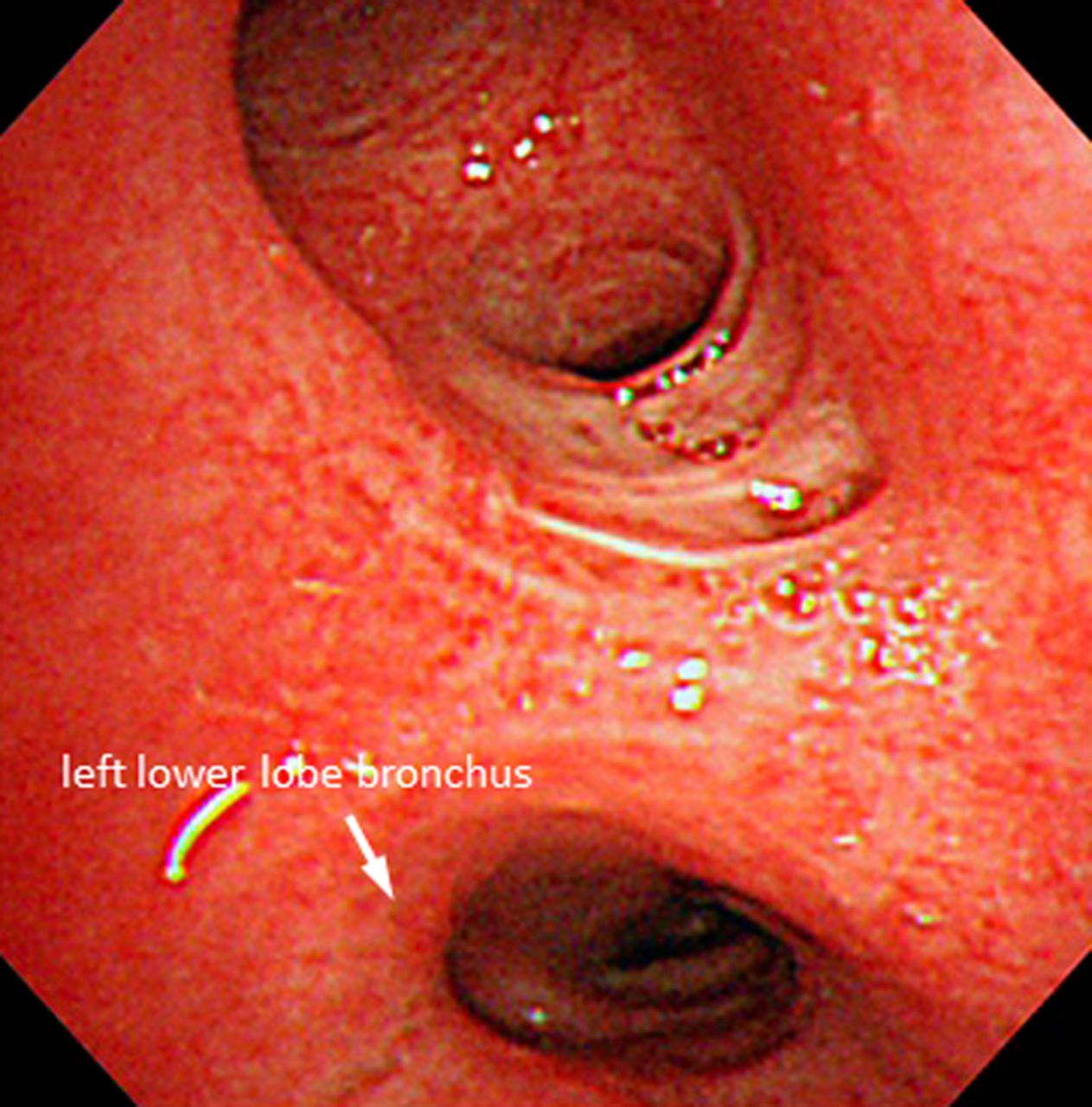
A bronchoscopy image showing diffuse redness and thickening of the bronchial mucosa with mild stenosis of the left lower bronchus.

A transbronchial biopsy showing neurinoma composed of spindle cells with a palisading pattern (H&E staining, ×400) containing S-100 protein-positive cells (inset).
Footnotes
-
Contributors HK is the doctor in charge of the patient, and wrote the first draft of the manuscript. HK and HI performed a transbronchial lung biopsy and rewrote new drafts based on comments from co-authors. FO provided radiological confirmation and constructed figures. All authors provided clinical, radiological and pathological confirmation of primary endobronchial neurinoma, and approved the final manuscript.
-
Competing interests None.
-
Patient consent Obtained.
-
Provenance and peer review Not commissioned; externally peer reviewed.