Article Text

Statistics from Altmetric.com
Description
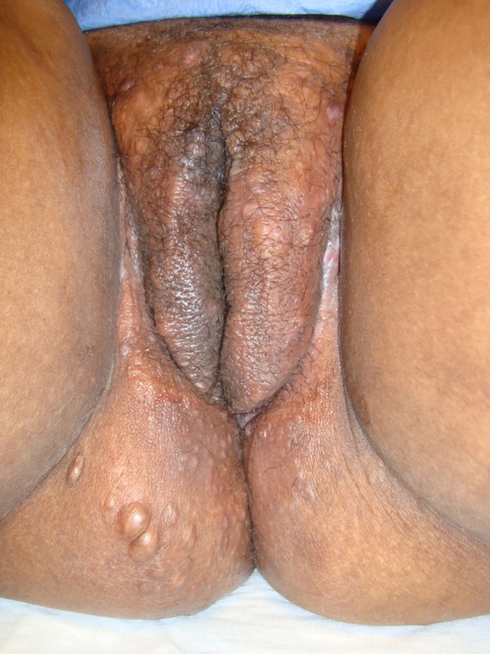
A 26-year-old woman was referred to dermatology department from an outside clinic for a 1-year history of recurrent draining lesions of the vulva and buttock treated unsuccessfully with doxycycline for presumed lymphogranuloma venereum. Physical examination was notable for an oedematous vulva (figure 1) with knife-like fissuring of the genitocrural folds (figure 2), superficial erosions and scattered draining papulonodules (figure 1). Biopsy showed granulomatous dermatitis consistent with cutaneous Crohn's disease. Colonoscopy revealed normal-appearing mucosa in the colon and terminal ileum, however, random biopsies demonstrated patchy, chronic inactive colitis with microgranulomas. MRI of the pelvis revealed multiple perivaginal and perirectal fistulas. The patient was initially managed with oral metronidazole and intralesional corticosteroid injections of the groin. She was started on adalimumab 40 mg. Her clinical course was complicated by recurrent perianal and axillary abscesses, requiring antibiotics, incision and drainage, and deferral of adalimumab, which was reinitiated following resolution of her active infections.

Oedematous vulva with superficial erosions and scattered papulonodules.

Knife-like fissuring of the genitocrural folds.
Cutaneous Crohn's manifesting with vulvar lesions is an under-recognised condition, with only 101 cases having been reported since 1965.1 The most frequently observed lesions include vulvar oedema (figure 1), ‘knife-like’ ulcerations (figure 2), hypertrophic lesions and chronic suppuration. Cutaneous Crohn's should be included on the differential diagnosis of vulvar lesions as these lesions may precede the diagnosis of gastrointestinal disease in 25% of cases.2 The differential diagnosis includes infections, lymphogranuloma venereum, granuloma inguinale, hidradenitis suppurativa and syphilis. Common treatments include oral steroids, metronidazole, azathioprine and 5-aminosalicylates. In addition, several case reports have documented successfully treated cutaneous Crohn's disease with either infliximab or adalimumab in patients who have failed other systemic immunosuppressive therapy.3
Learning points
-
Cutaneous Crohn's disease may present in patients without gastrointestinal symptoms.
-
Vulvar lesions of cutaneous Crohn's are classically linear ‘knife-like’ ulcerations along the genitocrural folds and vulvar oedema.
-
Treatment of cutaneous Crohn's disease requires a multidisciplinary approach, and while no specific treatment guidelines exist antitumour necrosis factor therapy may be a promising treatment option for control of this disease.
Footnotes
-
Contributors All the authors were involved with the clinical care of the patient. DD conducted literature search and initial drafting of the manuscript. MLS developed the idea for the article and was involved with the drafting of the manuscript. LBM and MKP performed the final editing of the manuscript. MKP provided the photographs.
-
Competing interests None.
-
Patient consent Obtained.
-
Provenance and peer review Not commissioned; externally peer reviewed.